 Explore
 Validate
 Learn
Learn Western blot
Antibody data
- Antibody Data
- Antigen structure
- References [0]
- Comments [0]
- Validations
- Western blot [2]
- Immunocytochemistry [2]
- Immunohistochemistry [1]
Submit
Validation data
Reference
Comment
Report error
- Product number
- 702787 - Provider product page

- Provider
- Invitrogen Antibodies
- Product name
- VAMP1 Recombinant Rabbit Monoclonal Antibody (24H9L10)
- Antibody type
- Monoclonal
- Antigen
- Synthetic peptide
- Description
- This antibody is predicted to react with Monkey, Mouse, Pig, Rabbit, Bovine.
- Antibody clone number
- 24H9L10
- Concentration
- 0.5 mg/mL
No comments: Submit comment
Supportive validation
- Submitted by
- Invitrogen Antibodies (provider)
- Main image
- Experimental details
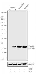
- Western blot analysis was performed on Membrane enriched extracts (30 µg lysate) of PC-12 (Lane 1), PC-12 treated with NGF and KCl (NGF at 200 nM, KCl at 50 nM for 7 days) (Lane 2) and tissue extracts (30 µg lysate) of Mouse Brain (Lane 3) and Rat Brain (Lane 4). The blots were probed with Anti-VAMP1 Recombinant Rabbit Monoclonal Antibody (Product # 702787, 2.5 µg/mL) and detected by chemiluminescence using Goat anti-Rabbit IgG (H+L) Superclonal™ Secondary Antibody, HRP conjugate (Product # A27036, 0.25 µg/mL, 1:4000 dilution). A 16 kDa band corresponding to VAMP1 was observed specifically in NGF differentiated PC-12 cells and tissues tested. Known quantity of protein samples were electrophoresed using Novex®NuPAGE®10% Bis-Tris gel (Product # NP0301BOX), XCell SureLock™ Electrophoresis System (Product # EI0002) and Novex® Sharp Pre-Stained Protein Standard (Product # LC5800). Resolved proteins were then transferred onto a nitrocellulose membrane with iBlot® Dry Blotting System (Product # IB21001). The membrane was probed with the relevant primary and secondary Antibody following blocking with 5% skimmed milk. Chemiluminescent detection was performed using Pierce™ ECL Western blotting Substrate (Product # 32106).

- Submitted by
- Invitrogen Antibodies (provider)
- Main image

- Experimental details
- Western blot analysis was performed on Membrane enriched extracts (30 µg lysate) of PC-12 (Lane 1), PC-12 treated with NGF and KCl (NGF at 200 nM, KCl at 50 nM for 7 days) (Lane 2) and tissue extracts (30 µg lysate) of Mouse Brain (Lane 3) and Rat Brain (Lane 4). The blots were probed with Anti-VAMP1 Recombinant Rabbit Monoclonal Antibody (Product # 702787, 2.5 µg/mL) and detected by chemiluminescence using Goat anti-Rabbit IgG (H+L) Superclonal™ Secondary Antibody, HRP conjugate (Product # A27036, 0.25 µg/mL, 1:4000 dilution). A 16 kDa band corresponding to VAMP1 was observed specifically in NGF differentiated PC-12 cells and tissues tested. Known quantity of protein samples were electrophoresed using Novex®NuPAGE®10% Bis-Tris gel (Product # NP0301BOX), XCell SureLock™ Electrophoresis System (Product # EI0002) and Novex® Sharp Pre-Stained Protein Standard (Product # LC5800). Resolved proteins were then transferred onto a nitrocellulose membrane with iBlot® Dry Blotting System (Product # IB21001). The membrane was probed with the relevant primary and secondary Antibody following blocking with 5% skimmed milk. Chemiluminescent detection was performed using Pierce™ ECL Western blotting Substrate (Product # 32106).

Supportive validation
- Submitted by
- Invitrogen Antibodies (provider)
- Main image
- Experimental details
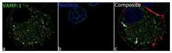
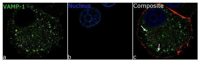
- For immunofluorescence analysis, PC12 cells were fixed and permeabilized for detection of endogenous VAMP1 using Anti- VAMP1 Recombinant Rabbit Monoclonal Antibody (Product # 702787, 5 µg/mL) and labeled with Goat anti-Rabbit IgG (H+L) Superclonal™ Secondary Antibody, Alexa Fluor® 488 conjugate (Product # A27034, 1:2000). Panel a) shows representative cells that were stained for detection and localization of VAMP1 protein (green), Panel b) is stained for nuclei (blue) using SlowFade® Gold Antifade Mountant with DAPI (Product # S36938). anel c) is a composite image of Panels a and b clearly demonstrating localisation of VAMP-1 in cytoplamic vesicles. The images were captured at 100X magnification.
- Submitted by
- Invitrogen Antibodies (provider)
- Main image

- Experimental details
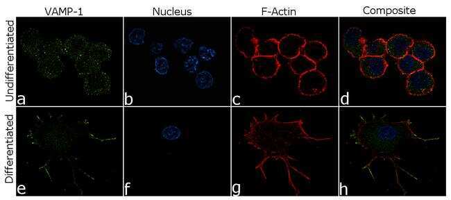
- For immunofluorescence analysis, PC12 cells were fixed and permeabilized for detection of endogenous VAMP1 using Anti-VAMP1 Recombinant Rabbit Monoclonal Antibody (Product # 702787, 5 µg/mL) and labeled with Goat anti-Rabbit IgG (H+L) Superclonal™ Secondary Antibody, Alexa Fluor® 488 conjugate (Product # A27034, 1:2000). Nuclei (blue) is stained using SlowFade® Gold Antifade Mountant with DAPI (Product # S36938) and cytoskeletal F-actin (red) staining using Rhodamine Phalloidin (Product # R415, 1:300) Panel a-d) shows representative undifferentiated cells that were stained for detection and localization of VAMP1 protein (green) with no signal. Panel e-h) clearly demonstrate enhanced signal of VMAP1 in cell body and neuronal processes of NGF (200 ng/mL 5days) induced differentiated PC12 cells. The images were captured at 60X magnification.
Supportive validation
- Submitted by
- Invitrogen Antibodies (provider)
- Main image
- Experimental details
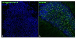
- Immunofluorescence analysis of mouse brain tissue: Frozen sections were fixed with 4% PFA for 20 min, permeabilized using 0.1% Triton-X 100 for 10 mins and blocked for 1 hour with 2% BSA. Transverse sections of mouse brain were incubated with Anti-VAMP1 Recombinant Rabbit Polyclonal Antibody (Product # 702787, 1:250 dilution) overnight at 4°C, followed by Goat anti-Rabbit IgG (H+L) Superclonal™ Secondary Antibody, Alexa Fluor® 488 conjugate (Product # A27034, 1:2000, 45 mins). Nuclei (blue) were stained using SlowFade® Gold Antifade Mountant with DAPI (Product # S36938), Panel a) represents staining with the matched isotype control. Panel b) shows a representative hippocampal region stained for VAMP1 (green). The images were captured at 20X magnification.
