 Explore
 Validate
 Learn
Learn Western blot
 ELISA
ELISA Immunohistochemistry
Antibody data
- Antibody Data
- Antigen structure
- References [0]
- Comments [0]
- Validations
- Immunohistochemistry [1]
Submit
Validation data
Reference
Comment
Report error
- Product number
- 200-301-G63 - Provider product page

- Provider
- Rockland Immunochemicals, Inc.
- Proper citation
- Rockland Cat#200-301-G63, RRID:AB_2611259
- Product name
- Anti-VAMP 1 and 2 (MOUSE) Monoclonal Antibody - 200-301-G63
- Antibody type
- Monoclonal
- Antigen
- Other
- Description
- Anti-VAMP 1 and 2 Antibody was purified by Protein G chromatography. This monoclonal antibody is specific for human synaptic protein. A BLAST analysis was used to suggest cross-reactivity with VAMP 1 and 2 from human based on 100% homology with the immunizing sequence. Recognzies the human synaptic vesicle protein syanptobrevin, VAMP, Recognizes VAMP 1 and 2. Cross-reactivity with VAMP 1 and 2 from other sources has not been determined. Neurobiology research.
- Reactivity
- Human
- Host
- Mouse
- Conjugate
- Unconjugated
- Isotype
- IgG
- Antibody clone number
- SP-11
- Vial size
- 100 µl
- Concentration
- 1.0 mg/ml
- Storage
- Store vial at -20° C prior to opening. Aliquot contents and freeze at -20° C or below for extended storage. Avoid cycles of freezing and thawing. Centrifuge product if not completely clear after standing at room temperature. This product is stable for several weeks at 4° C as an undiluted liquid. Dilute only prior to immediate use.
- Handling
- Dry Ice
No comments: Submit comment
Supportive validation
- Submitted by
- Rockland Immunochemicals, Inc. (provider)
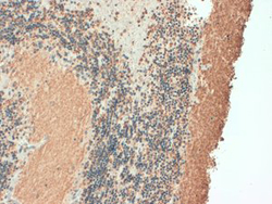
- Main image
- Experimental details
- Immunohistochemistry of mouse anti-VAMP1 AND 2 antibody. Tissue: Human Brain cerebellum neurons and fibers. Fixation: N/A. Antigen Retrieval: not required. Primary Antibody: Vamp1 and 2 antibody at 1 ?g/mL for 1h at RT. Secondary antibody: Peroxidase mouse secondary at 1:10,000 for 45 min at RT. Localization: Cytoplasmic vesicle membrane. Staining: Vamp1 and 2 as brown signal.
- Validation comment
- Immunohistochemistry
