Explore
 Validate
 Learn
Learn Western blot
Antibody data
- Antibody Data
- Antigen structure
- References [4]
- Comments [0]
- Validations
- Western blot [3]
- Immunoprecipitation [1]
- Immunohistochemistry [4]
Submit
Validation data
Reference
Comment
Report error
- Product number
- NB100-40840 - Provider product page

- Provider
- Novus Biologicals
- Proper citation
- Novus Cat#NB100-40840, RRID:AB_789689
- Product name
- Rabbit Polyclonal CDT2 Antibody
- Antibody type
- Polyclonal
- Description
- Immunogen affinity purified.
- Reactivity
- Human, Mouse
- Host
- Rabbit
- Isotype
- IgG
- Vial size
- 0.1 ml
- Concentration
- 0.2 mg/ml
- Storage
- Store at 4C. Do not freeze.
Submitted references Overexpression of denticleless E3 ubiquitin protein ligase homolog (DTL) is related to poor outcome in gastric carcinoma.
Tumor suppressor protein Lgl mediates G1 cell cycle arrest at high cell density by forming an Lgl-VprBP-DDB1 complex.
Regulation of the CRL4(Cdt2) ubiquitin ligase and cell-cycle exit by the SCF(Fbxo11) ubiquitin ligase.
SET8 is degraded via PCNA-coupled CRL4(CDT2) ubiquitylation in S phase and after UV irradiation.
Kobayashi H, Komatsu S, Ichikawa D, Kawaguchi T, Hirajima S, Miyamae M, Okajima W, Ohashi T, Kosuga T, Konishi H, Shiozaki A, Fujiwara H, Okamoto K, Tsuda H, Otsuji E
Oncotarget 2015 Nov 3;6(34):36615-24
Oncotarget 2015 Nov 3;6(34):36615-24
Tumor suppressor protein Lgl mediates G1 cell cycle arrest at high cell density by forming an Lgl-VprBP-DDB1 complex.
Yamashita K, Ide M, Furukawa KT, Suzuki A, Hirano H, Ohno S
Molecular biology of the cell 2015 Jul 1;26(13):2426-38
Molecular biology of the cell 2015 Jul 1;26(13):2426-38
Regulation of the CRL4(Cdt2) ubiquitin ligase and cell-cycle exit by the SCF(Fbxo11) ubiquitin ligase.
Rossi M, Duan S, Jeong YT, Horn M, Saraf A, Florens L, Washburn MP, Antebi A, Pagano M
Molecular cell 2013 Mar 28;49(6):1159-66
Molecular cell 2013 Mar 28;49(6):1159-66
SET8 is degraded via PCNA-coupled CRL4(CDT2) ubiquitylation in S phase and after UV irradiation.
Jørgensen S, Eskildsen M, Fugger K, Hansen L, Larsen MS, Kousholt AN, Syljuåsen RG, Trelle MB, Jensen ON, Helin K, Sørensen CS
The Journal of cell biology 2011 Jan 10;192(1):43-54
The Journal of cell biology 2011 Jan 10;192(1):43-54
No comments: Submit comment
Supportive validation
- Submitted by
- Novus Biologicals (provider)
- Main image
- Experimental details
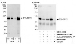
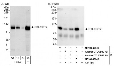
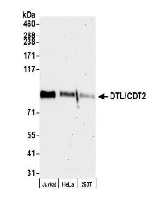
- Western Blot: CDT2 Antibody [NB100-40840] - Detection of Human DTL/CDT2 by Western Blot and Immunoprecipitation. Samples: Whole cell lysate from HeLa (5, 15 and 50 ug for WB; 1 mg for IP, 20% of IP loaded) and 293T (T; 50 ug) cells. Antibodies: Affinity purified rabbit anti- DTL/CDT2 antibody NB100-40840 used for WB at 0.04 ug/ml (A) and 1 ug/ml (B) and used for IP at 3 ug/mg lysate (B). DTL/CDT2 was also immunoprecipitated by other rabbit anti- DTL/CDT2 antibodies and NB100-40839, which recognize upstream epitopes. Detection: Chemiluminescence with exposure times of 30 seconds (A) and 10 seconds (B).
- Submitted by
- Novus Biologicals (provider)
- Main image
- Experimental details
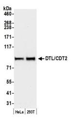
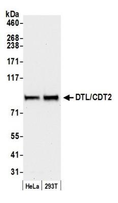
- Western Blot: CDT2 Antibody [NB100-40840] - Detection of Human DTL/CDT2 by Western Blot. Samples: Whole cell lysate (50 ug) from HeLa and 293T cells prepared using NETN lysis buffer. Antibody: Affinity purified rabbit anti-DTL/CDT2 antibody NB100-40840 used for WB at 0.1 ug/ml. Detection: Chemiluminescence with an exposure time of 10 seconds.
- Submitted by
- Novus Biologicals (provider)
- Main image
- Experimental details
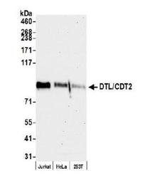
- Western Blot: CDT2 Antibody [NB100-40840] - Whole cell lysate (15 ug) from Jurkat, HeLa, and HEK293T cells prepared using NETN lysis buffer. Antibody: Affinity purified rabbit anti-DTL/CDT2 antibody used for WB at 0.04 ug/ml. Detection: Chemiluminescence with an exposure time of 75 seconds.
Supportive validation
- Submitted by
- Novus Biologicals (provider)
- Main image
- Experimental details
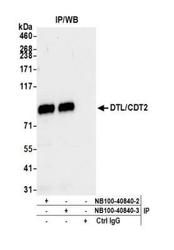
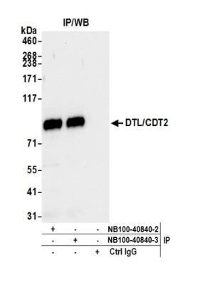
- Immunoprecipitation: CDT2 Antibody [NB100-40840] - Detection of human DTL/CDT2 by western blot of immunoprecipitates. Samples: Whole cell lysate (1.0 mg per IP reaction; 20% of IP loaded) from Jurkat cells prepared using NETN lysis buffer. Antibodies: Affinity purified rabbit anti-DTL/CDT2 antibody NB100-40840 (lot NB100-40840-3) used for IP at 6 ug per reaction. DTL/CDT2 was also immunoprecipitated by a previous lot of this antibody (lot NB100-40840-2). For blotting immunoprecipitated DTL/CDT2, NB100-40840 was used at 0.1 ug/ml. Detection: Chemiluminescence with an exposure time of 3 seconds.
Supportive validation
- Submitted by
- Novus Biologicals (provider)
- Main image
- Experimental details
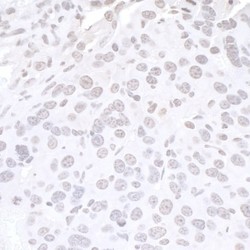
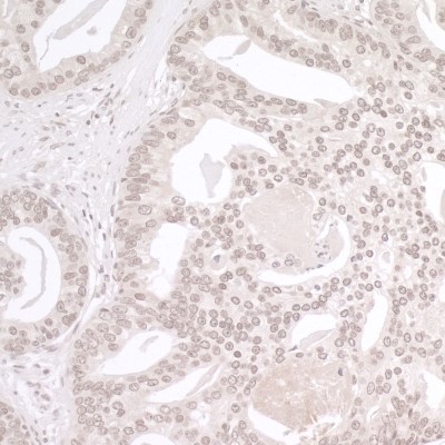
- Immunohistochemistry: CDT2 Antibody [NB100-40840] - Sample: FFPE section of mouse renal cell carcinoma. Antibody: Affinity purified rabbit anti-DTL/CDT2 used at a dilution of 1:200 (1ug/ml). Detection: DAB
- Submitted by
- Novus Biologicals (provider)
- Main image
- Experimental details
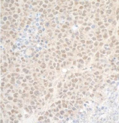
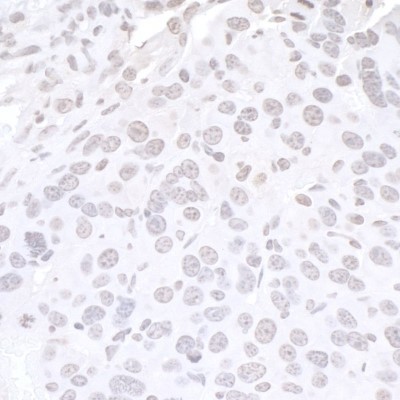
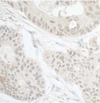
- Immunohistochemistry: CDT2 Antibody [NB100-40840] - Sample: FFPE section of human prostate carcinoma. Antibody: Affinity purified rabbit anti-DTL/CDT2 used at a dilution of 1:200 (1ug/ml). Detection: DAB
- Submitted by
- Novus Biologicals (provider)
- Main image

- Experimental details
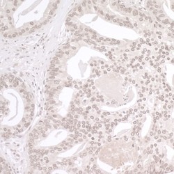
- Immunohistochemistry-Paraffin: CDT2 Antibody [NB100-40840] - Section of human prostate carcinoma. Antibody: Affinity purified rabbit anti- DTL/CDT2 used at a dilution of 1:1000 (0.2 ug/ml). Detection: DAB
- Submitted by
- Novus Biologicals (provider)
- Main image
- Experimental details
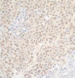
- Immunohistochemistry-Paraffin: CDT2 Antibody [NB100-40840] - Section of mouse plasmacytoma. Antibody: Affinity purified rabbit anti-DTL/CDT2 used at a dilution of 1:200 (1ug/ml). Detection: DAB