 Explore
 Validate
 Learn
Learn Western blot
Antibody data
- Antibody Data
- Antigen structure
- References [0]
- Comments [0]
- Validations
- Western blot [4]
- Immunocytochemistry [1]
- Immunoprecipitation [1]
- Immunohistochemistry [2]
Submit
Validation data
Reference
Comment
Report error
- Product number
- GTX103235 - Provider product page

- Provider
- GeneTex
- Proper citation
- GeneTex Cat#GTX103235, RRID:AB_11179576
- Product name
- Thymidylate synthase antibody
- Antibody type
- Polyclonal
- Reactivity
- Human, Mouse, Rat
- Host
- Rabbit
No comments: Submit comment
Supportive validation
- Submitted by
- GeneTex (provider)
- Main image

- Experimental details
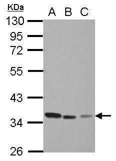
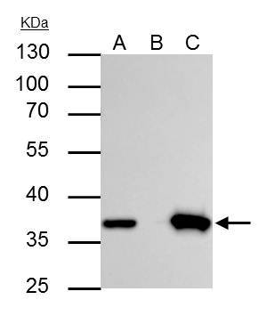
- Sample (30 ug of whole cell lysate) A: Jurkat B: Raji C: THP-1 10% SDS PAGE GTX103235 diluted at 1:1000
- Submitted by
- GeneTex (provider)
- Main image
- Experimental details
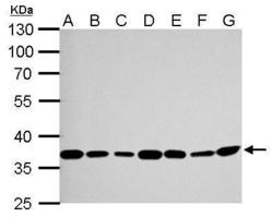
- Thymidylate synthetase antibody detects TYMS protein by Western blot analysis.A. 30 ?g Neuro2A whole cell lysate/extract B. 30 ?g GL261 whole cell lysate/extract C. 30 ?g C8D30 whole cell lysate/extract D. 30 ?g NIH-3T3 whole cell lysate/extract E. 30 ?g BCL-1 whole cell lysate/extract F. 30 ?g Raw264.7 whole cell lysate/extract G. 30 ?g C2C12 whole cell lysate/extract 10 % SDS-PAGEThymidylate synthetase antibody (GTX103235) dilution: 1:1000

- Submitted by
- GeneTex (provider)
- Main image

- Experimental details
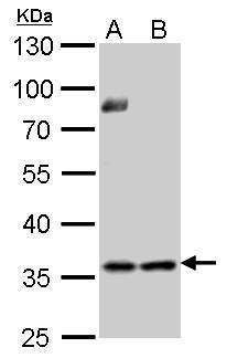
- Thymidylate synthetase antibody detects TYMS protein by Western blot analysis.A. 30 ?g PC-12 whole cell lysate/extract B. 30 ?g Rat2 whole cell lysate/extract10 % SDS-PAGEThymidylate synthetase antibody (GTX103235) dilution: 1:1000
- Submitted by
- GeneTex (provider)
- Main image

- Experimental details
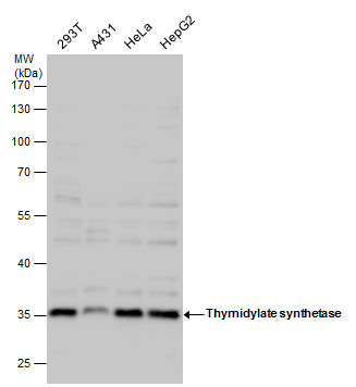
- Thymidylate synthetase antibody detects Thymidylate synthetase protein by western blot analysis. Various whole cell extracts (30 £gg) were separated by 10% SDS-PAGE, and the membrane was blotted with Thymidylate synthetase antibody (GTX103235) diluted by 1:1000.
Supportive validation
- Submitted by
- GeneTex (provider)
- Main image

- Experimental details
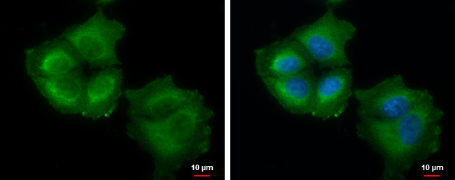
- Thymidylate synthetase antibody detects Thymidylate synthetase protein at cytoplasm by immunofluorescent analysis.Sample: A375 cells were fixed in 2% paraformaldehyde/culture medium at 37¢J for 30 min.Green: Thymidylate synthetase protein stained by Thymidylate synthetase antibody (GTX103235) diluted at 1:1000.Blue: Hoechst 33342 staining.
Supportive validation
- Submitted by
- GeneTex (provider)
- Main image

- Experimental details
- Thymidylate synthetase antibody immunoprecipitates TYMS protein in IP experiments. IP Sample: Jurkat whole cell lysate/extract A : 30 £gg whole cell lysate/extract of TYMS protein expressing Jurkat cells B : Control with 3 £gg of pre-immune rabbit IgG C : Immunoprecipitation of TYMS by 3 £gg of thymidylate synthetase antibody (GTX103235) 10% SDS-PAGE The immunoprecipitated TYMS protein was detected by thymidylate synthetase antibody (GTX103235) diluted at 1 : 1000. EasyBlot anti-rabbit IgG (HRP) (GTX221666-01) was used as a secondary reagent.
Supportive validation
- Submitted by
- GeneTex (provider)
- Main image

- Experimental details
- Thymidylate synthetase antibody detects Thymidylate synthetase protein at cytosol and nucleus on human ovarian carcinoma by immunohistochemical analysis. Sample: Paraffin-embedded human ovarian carcinoma. Thymidylate synthetase antibody (GTX103235) dilution: 1:500.

- Submitted by
- GeneTex (provider)
- Main image

- Experimental details
- Thymidylate synthetase antibody detects Thymidylate synthetase protein at cytosol and nucleus on human colon carcinoma by immunohistochemical analysis. Sample: Paraffin-embedded human colon carcinoma. Thymidylate synthetase antibody (GTX103235) dilution: 1:500.
