Explore
 Validate
 Learn
Learn Western blot
Antibody data
- Antibody Data
- Antigen structure
- References [2]
- Comments [0]
- Validations
- Western blot [1]
- Other assay [1]
Submit
Validation data
Reference
Comment
Report error
- Product number
- PA5-42709 - Provider product page

- Provider
- Invitrogen Antibodies
- Product name
- Lamin B Receptor Polyclonal Antibody
- Antibody type
- Polyclonal
- Antigen
- Synthetic peptide
- Description
- Peptide sequence: GANSQKNAFR KNPSDPKLAH LKTIHTSTGK NLLVSGWWGF VRHPNYLGDL Sequence homology: Cow: 86%; Dog: 93%; Guinea Pig: 86%; Horse: 93%; Human: 100%; Mouse: 93%; Pig: 93%; Rabbit: 93%; Rat: 100%; Yeast: 77%; Zebrafish: 79%
- Reactivity
- Human
- Host
- Rabbit
- Isotype
- IgG
- Vial size
- 100 µL
- Concentration
- 0.5 mg/mL
- Storage
- -20° C, Avoid Freeze/Thaw Cycles
Submitted references Expression of myelin transcription factor 1 and lamin B receptor mediate neural progenitor fate transition in the zebrafish spinal cord pMN domain.
Alterations in nuclear structure promote lupus autoimmunity in a mouse model.
Xing L, Chai R, Wang J, Lin J, Li H, Wang Y, Lai B, Sun J, Chen G
The Journal of biological chemistry 2022 Oct;298(10):102452
The Journal of biological chemistry 2022 Oct;298(10):102452
Alterations in nuclear structure promote lupus autoimmunity in a mouse model.
Singh N, Johnstone DB, Martin KA, Tempera I, Kaplan MJ, Denny MF
Disease models & mechanisms 2016 Aug 1;9(8):885-97
Disease models & mechanisms 2016 Aug 1;9(8):885-97
No comments: Submit comment
Supportive validation
- Submitted by
- Invitrogen Antibodies (provider)
- Main image
- Experimental details
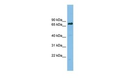
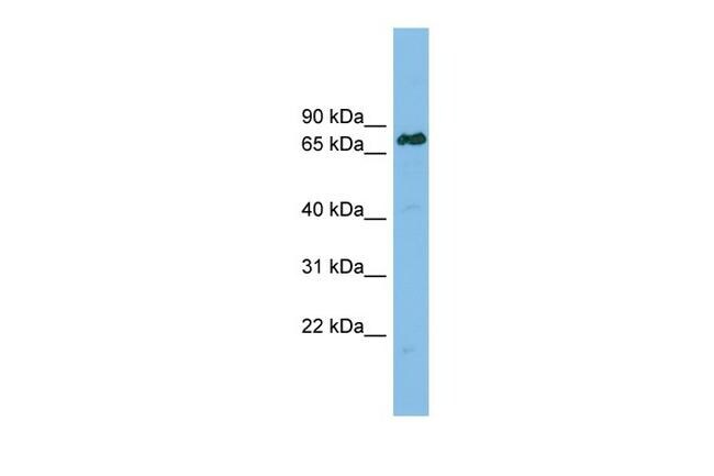
- Western blot analysis of human RPMI 8226 cell lysate using an anti-Lamin B Receptor polyclonal antibody (Product # PA5-42709).
Supportive validation
- Submitted by
- Invitrogen Antibodies (provider)
- Main image

- Experimental details
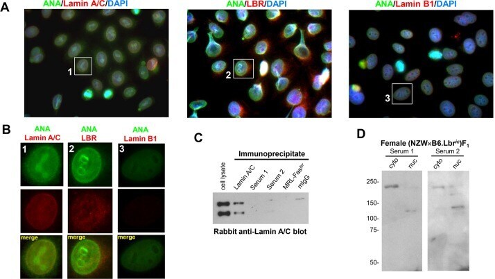
- Fig. 5. Female (NZWxB6.Lbr ic )F 1 mice develop anti-nuclear autoantibodies recognizing the A-type lamina. (A) Co-staining of HEp-2 cells with female (NZWxB6.Lbr ic )F 1 serum and lamin A/C (left panel), LBR (middle panel), and lamin B1 (right panel). Anti-nuclear antibody (ANA) staining in the serum was detected with an Alexa Fluor 488-conjugated goat anti-mouse antibody, lamina proteins were labeled with the indicated rabbit anti-serum and an Alexa Fluor 594-conjugated goat anti-rabbit secondary antibody. Nuclei were counterstained with DAPI. Image magnification, 1000x. The individual red, green and blue color channels demonstrating the co-localization of the nuclear membrane component of anti-nuclear autoantibody reactivity with the A-type lamina are presented in Fig. S6 . (B) Digital enlargement of the individual cells indicated in A. Anti-nuclear antibody staining mediated by the (NZWxB6.Lbr ic )F 1 serum is shown in the green channel, and the indicated nuclear envelope protein is counterstained in the red channel. The nuclear membrane colocalization of mouse serum with the anti-lamin A/C is represented by the yellow overlap signal. (C) Lamin A/C immunoblots of 5x10 6 MEFs immunoprecipitated with a mouse lamin A/C monoclonal antibody, the sera from two female (NZWxB6.Lbr ic )F 1 mice (Serum 1 and 2), or pooled aged female MRL-Fas lpr sera. Non-specific mouse IgG (mIgG) served as a serum control. (D) Cytoplasmic and nuclear extracts prepared from 1x10 6 MEFs were resolved