 Explore
 Validate
 Learn
Learn Western blot
 Immunohistochemistry
Antibody data
- Antibody Data
- Antigen structure
- References [0]
- Comments [0]
- Validations
- Western blot [4]
- Immunocytochemistry [2]
- Immunohistochemistry [1]
Submit
Validation data
Reference
Comment
Report error
- Product number
- HPA027815 - Provider product page

- Provider
- Atlas Antibodies
- Proper citation
- Atlas Antibodies Cat#HPA027815, RRID:AB_10609994
- Product name
- Anti-IRF2BP2
- Antibody type
- Polyclonal
- Reactivity
- Human, Mouse, Rat
- Host
- Rabbit
- Conjugate
- Unconjugated
- Antigen sequence
QDWVNRPKTVRDTLLALHQHGHSGPFESKFKKEPA
LTAGRLLGFEANGANGSKAVARTARKRKPSPEPEG
EVG- Isotype
- IgG
- Vial size
- 100 µl
- Storage
- Store at +4°C for short term storage. Long time storage is recommended at -20°C.
No comments: Submit comment
Enhanced validation
Supportive validation
- Submitted by
- klas2
- Enhanced method
- Genetic validation
- Main image
- Experimental details
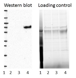
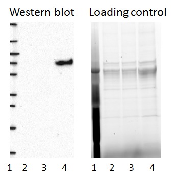
- Western blot of cell lysate from U-2 OS cells transfected with either siRNA targeting IRF2BP2 or control siRNA. Lane 1: Marker (250, 130, 95, 72, 55, 36, 28, 17, 10) Lane 2: Cell lysate from U-2OS cells transfected with siRNA targeting IRF2BP2 Lane 3: N/A Lane 4: Cell lysate from U-2OS cells transfected with control siRNA Right image, lane 1-4: loading control
- Sample type
- U-2 OS
- Primary Ab dilution
- 1:219
- Conjugate
- Horseradish Peroxidase
- Secondary Ab
- Secondary Ab
- Secondary Ab dilution
- 1:3000
- Knockdown/Genetic Approaches Application
- Western blot
Supportive validation
- Submitted by
- Atlas Antibodies (provider)
- Enhanced method
- Genetic validation
- Main image
- Experimental details
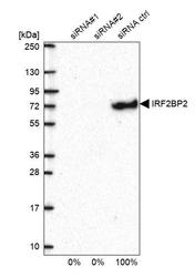
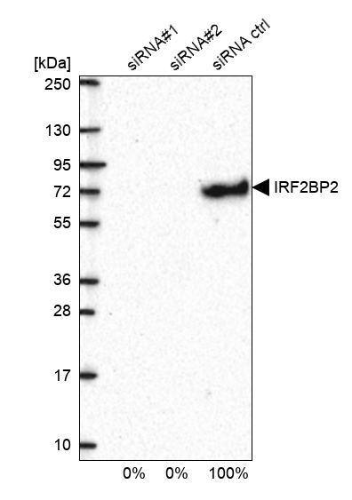
- Western blot analysis in U2OS cells transfected with control siRNA, target specific siRNA probe #1 and #2, using Anti-IRF2BP2 antibody. Remaining relative intensity is presented.
- Submitted by
- Atlas Antibodies (provider)
- Main image

- Experimental details
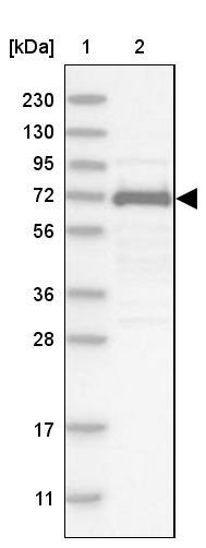
- Lane 1: Marker [kDa] 230, 130, 95, 72, 56, 36, 28, 17, 11Lane 2: Human cell line RT-4
- Submitted by
- Atlas Antibodies (provider)
- Main image

- Experimental details
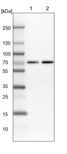
- Lane 1: NIH-3T3 cell lysate (Mouse embryonic fibroblast cells)Lane 2: NBT-II cell lysate (Rat Wistar bladder tumour cells)
Enhanced validation
Supportive validation
- Submitted by
- 55af80e3e0991
- Enhanced method
- Genetic validation
- Main image
- Experimental details
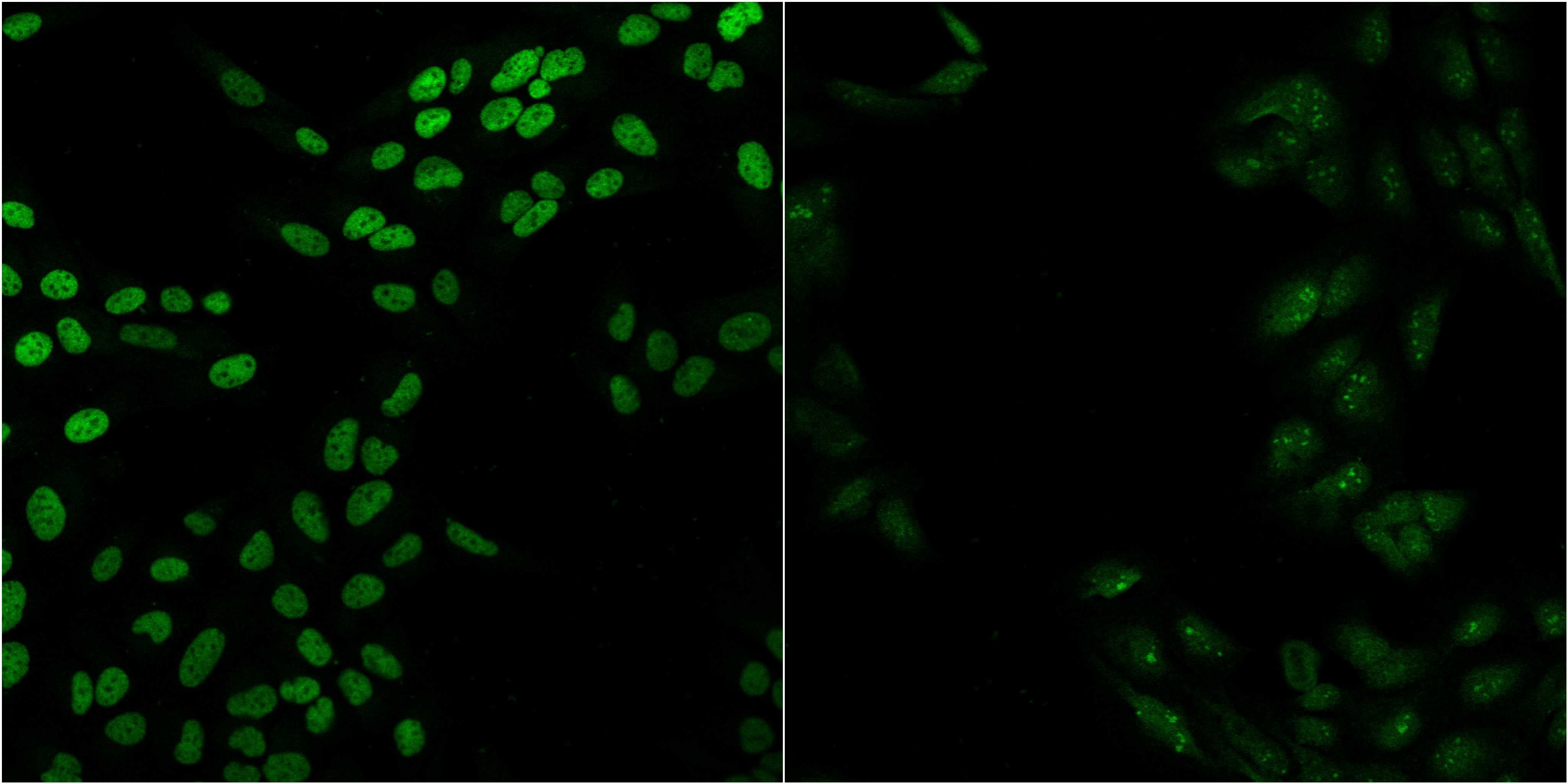
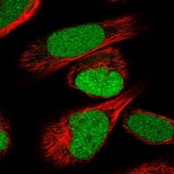
- Confocal images of immunofluorescently stained human U-2 OS cells.The protein IRF2BP2 is shown in green. The image to the left show cells transfected with control siRNA and the image to the right show cells where IRF2BP2 has been downregulated with specific siRNA.
- Sample type
- U-2 OS cells
- Primary Ab dilution
- 1:98
- Secondary Ab
- Secondary Ab
- Secondary Ab dilution
- 1:800
- Knockdown/Genetic Approaches Application
- Immunocytochemistry
Supportive validation
- Submitted by
- Atlas Antibodies (provider)
- Main image
- Experimental details
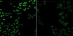
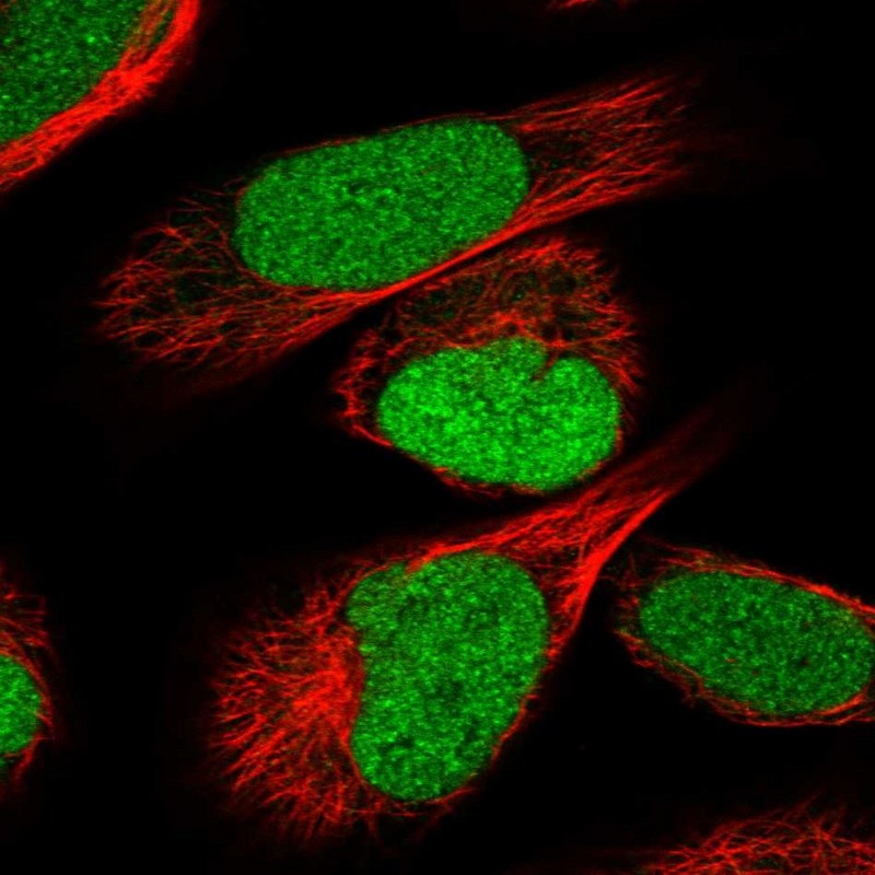
- Immunofluorescent staining of human cell line U-2 OS shows localization to nucleus.
- Sample type
- HUMAN
Supportive validation
- Submitted by
- Atlas Antibodies (provider)
- Main image
- Experimental details
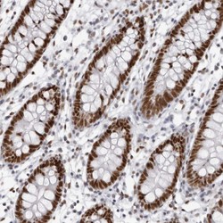
- Immunohistochemical staining of human colon shows strong nuclear positivity in glandular cells.
- Sample type
- HUMAN
