 Explore
 Validate
 Learn
Learn Western blot
Antibody data
- Antibody Data
- Antigen structure
- References [0]
- Comments [0]
- Validations
- Western blot [2]
- Immunocytochemistry [2]
- Immunohistochemistry [1]
- Flow cytometry [1]
Submit
Validation data
Reference
Comment
Report error
- Product number
- APR-022-25UL - Provider product page

- Provider
- Invitrogen Antibodies
- Product name
- P2X1 Receptor (extracellular) Polyclonal Antibody
- Antibody type
- Polyclonal
- Antigen
- Other
- Reactivity
- Human, Mouse, Rat
- Host
- Rabbit
- Isotype
- IgG
- Vial size
- 25 µL
- Concentration
- 0.8 mg/mL
- Storage
- -20° C, Avoid Freeze/Thaw Cycles
No comments: Submit comment
Supportive validation
- Submitted by
- Invitrogen Antibodies (provider)
- Main image
- Experimental details
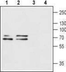
- Western blot analysis of rat brain (lanes 1 and 3) and mouse brain (lanes 2 and 4) lysates: - 1,2. Anti-P2X1 Receptor (extracellular) Antibody (#APR-022), (1:200).3,4. Anti-P2X1 Receptor (extracellular) Antibody , preincubated with P2X1 Receptor (extracellular) Blocking Peptide (#BLP-PR022).

- Submitted by
- Invitrogen Antibodies (provider)
- Main image

- Experimental details
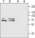
- Western blot analysis of rat brain (lanes 1 and 3) and mouse brain (lanes 2 and 4) lysates: - 1,2. Anti-P2X1 Receptor (extracellular) Antibody (#APR-022), (1:200).3,4. Anti-P2X1 Receptor (extracellular) Antibody , preincubated with P2X1 Receptor (extracellular) Blocking Peptide (#BLP-PR022).
Supportive validation
- Submitted by
- Invitrogen Antibodies (provider)
- Main image

- Experimental details
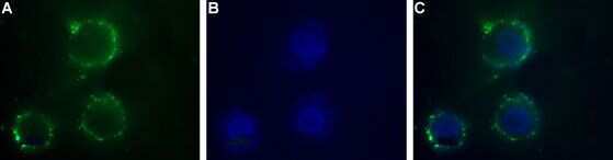
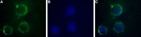
- Expression of P2X1 Receptor in human SH-SYS5 cells - Cell surface detection of P2X1 Receptor inintact livinghuman SH-SYS5 cells. A. Extracellular staining of cells with Anti-P2X1 Receptor (extracellular) Antibody (#APR-022), (1:50) followed by goat Anti-rabbit-AlexaFluor-488 secondary Antibody .B. Nuclear staining DAPI as the counterstain.C. Merged images of A and B.
- Submitted by
- Invitrogen Antibodies (provider)
- Main image
- Experimental details
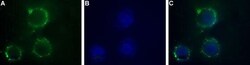
- Expression of P2X1 Receptor in human SH-SYS5 cells - Cell surface detection of P2X1 Receptor inintact livinghuman SH-SYS5 cells. A. Extracellular staining of cells with Anti-P2X1 Receptor (extracellular) Antibody (#APR-022), (1:50) followed by goat Anti-rabbit-AlexaFluor-488 secondary Antibody .B. Nuclear staining DAPI as the counterstain.C. Merged images of A and B.
Supportive validation
- Submitted by
- Invitrogen Antibodies (provider)
- Main image
- Experimental details
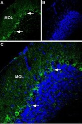
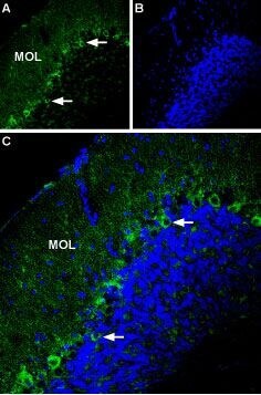
- Expression of P2X1 Receptor in mouse cerebellum - Immunohistochemical staining of mouse cerebellum using Anti-P2X1 Receptor (extracellular) Antibody (#APR-022). A. Most of P2RX1 labeling (green) appears in fine processes in the molecular layer (MOL) and in Purkinje cells (arrows show examples). B. DAPI is used as the counterstain (blue). C. Merge of A and B.
Supportive validation
- Submitted by
- Invitrogen Antibodies (provider)
- Main image
- Experimental details
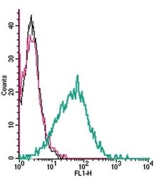
- Cell surface detection ofP2X1by indirect flow cytometry in live intacthumanMEG-01 megakaryocyticleukemiacells: - (black line) cells. (red) Cells+ goat- Anti-rabbit-FITC. (green) Cells+ Anti-P2X1 Receptor (extracellular) Antibody (#APR-022), (2.5μg) + goat- Anti-rabbit-FITC.
