Explore
 Validate
 Learn
Learn Western blot
Antibody data
- Antibody Data
- Antigen structure
- References [8]
- Comments [0]
- Validations
- Western blot [3]
- Immunocytochemistry [4]
- Immunohistochemistry [2]
- Other assay [1]
Submit
Validation data
Reference
Comment
Report error
- Product number
- MA1-752 - Provider product page

- Provider
- Invitrogen Antibodies
- Product name
- Presenilin 1 Monoclonal Antibody (APS 18)
- Antibody type
- Monoclonal
- Antigen
- Synthetic peptide
- Description
- MA1-752 detects presenilin 1 protein (PS1) from mouse, rat, human, and nonhuman primate samples. No cross-reactivity is seen with presenilin 2.
- Antibody clone number
- APS 18
- Concentration
- 1 mg/mL
Submitted references β-Secretase 1's Targeting Reduces Hyperphosphorilated Tau, Implying Autophagy Actors in 3xTg-AD Mice.
Self-renewal and differentiation of reactive astrocyte-derived neural stem/progenitor cells isolated from the cortical peri-infarct area after stroke.
Analysis of presenilin 1 and presenilin 2 expression and processing by newly developed monoclonal antibodies.
Analysis of presenilin 1 and presenilin 2 expression and processing by newly developed monoclonal antibodies.
Direct interaction of Alzheimer's disease-related presenilin 1 with armadillo protein p0071.
Direct interaction of Alzheimer's disease-related presenilin 1 with armadillo protein p0071.
Abrogation of the presenilin 1/beta-catenin interaction and preservation of the heterodimeric presenilin 1 complex following caspase activation.
Abrogation of the presenilin 1/beta-catenin interaction and preservation of the heterodimeric presenilin 1 complex following caspase activation.
Piedrahita D, Castro-Alvarez JF, Boudreau RL, Villegas-Lanau A, Kosik KS, Gallego-Gomez JC, Cardona-Gómez GP
Frontiers in cellular neuroscience 2015;9:498
Frontiers in cellular neuroscience 2015;9:498
Self-renewal and differentiation of reactive astrocyte-derived neural stem/progenitor cells isolated from the cortical peri-infarct area after stroke.
Shimada IS, LeComte MD, Granger JC, Quinlan NJ, Spees JL
The Journal of neuroscience : the official journal of the Society for Neuroscience 2012 Jun 6;32(23):7926-40
The Journal of neuroscience : the official journal of the Society for Neuroscience 2012 Jun 6;32(23):7926-40
Analysis of presenilin 1 and presenilin 2 expression and processing by newly developed monoclonal antibodies.
Diehlmann A, Ida N, Weggen S, Grünberg J, Haass C, Masters CL, Bayer TA, Beyreuther K
Journal of neuroscience research 1999 May 15;56(4):405-19
Journal of neuroscience research 1999 May 15;56(4):405-19
Direct interaction of Alzheimer's disease-related presenilin 1 with armadillo protein p0071.
Stahl B, Diehlmann A, Südhof TC
The Journal of biological chemistry 1999 Apr 2;274(14):9141-8
The Journal of biological chemistry 1999 Apr 2;274(14):9141-8
Direct interaction of Alzheimer's disease-related presenilin 1 with armadillo protein p0071.
Stahl B, Diehlmann A, Südhof TC
The Journal of biological chemistry 1999 Apr 2;274(14):9141-8
The Journal of biological chemistry 1999 Apr 2;274(14):9141-8
Abrogation of the presenilin 1/beta-catenin interaction and preservation of the heterodimeric presenilin 1 complex following caspase activation.
Tesco G, Kim TW, Diehlmann A, Beyreuther K, Tanzi RE
The Journal of biological chemistry 1998 Dec 18;273(51):33909-14
The Journal of biological chemistry 1998 Dec 18;273(51):33909-14
No comments: Submit comment
Supportive validation
- Submitted by
- Invitrogen Antibodies (provider)
- Main image

- Experimental details
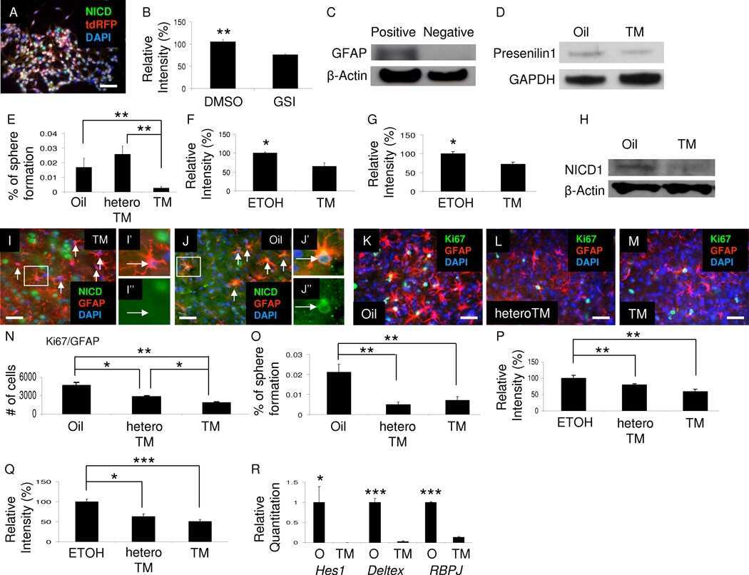
- Western blot analysis of Presenilin 1 was performed by loading 20 µg of THP-1 cell lysate per well onto an SDS-PAGE gel. Proteins were transferred to a PVDF membrane and blocked with 5% non-fat milk in TBST for 1 hour at room temperature. The membrane was probed with a Presenilin 1 monoclonal antibody (Product # MA1-752) at a dilution of 1:250 overnight at 4°C, washed in TBST, and probed with an HRP-conjugated goat anti-mouse IgG secondary antibody at a dilution of 1:40,000 for 1 hour at room temperature. Chemiluminescent detection was performed using ECL substrate. Data courtesy of the Innovators Program.
- Submitted by
- Invitrogen Antibodies (provider)
- Main image

- Experimental details
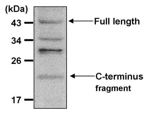
- Knockdown of Presenilin 1 was achieved by transfecting T-47D with Presenilin 1 specific siRNAs (Silencer® select Product # s112). Western blot analysis (Fig. a) was performed using Membrane enriched extracts from the Presenilin 1 knockdown cells (lane 3), non-targeting scrambled siRNA transfected cells (lane 2) and untransfected cells (lane 1). The blot was probed with Presenilin 1 Monoclonal Antibody (APS 18) (Product # MA1-752, 1:500 dilution ) and Goat anti-Mouse IgG (H+L) Superclonal™ Recombinant Secondary Antibody, HRP (Product # A28177, 1:4000 dilution). Densitometric analysis of this western blot is shown in histogram (Fig. b). Decrease in signal upon siRNA mediated knock down confirms that antibody is specific to Presenilin 1 C- terminal fragment.

- Submitted by
- Invitrogen Antibodies (provider)
- Main image
- Experimental details
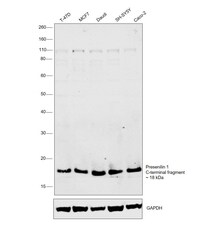
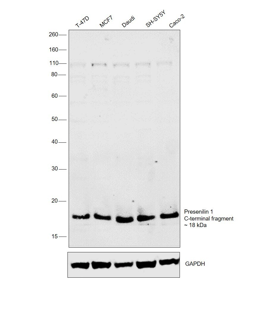
- Western blot was performed using Anti-Presenilin 1 Monoclonal Antibody (APS 18)(Product # MA1-752) and a 18kDa band corresponding to Presenilin 1 was observed across the cell lines tested. 18 kDa corresponds to the C-terminal fragment of Presenilin 1. Membrane enriched extracts (30 µg lysate) of T-47D (Lane 1), MCF7 (Lane 2), Daudi (Lane 3), SH-SY5Y (Lane 4), Caco-2 (Lane 5) were electrophoresed using NuPAGE™ 10% Bis-Tris Protein Gel (Product # NP0301BOX). Resolved proteins were then transferred onto a Nitrocellulose membrane (Product # IB23001) by iBlot® 2 Dry Blotting System (Product # IB21001). The blot was probed with the primary antibody (1:500 dilution) and detected by chemiluminescence with Goat anti-Mouse IgG (H+L) Superclonal™ Recombinant Secondary Antibody, HRP (Product # A28177,1:4000 dilution) using the iBright FL 1000 (Product # A32752). Chemiluminescent detection was performed using SuperSignal™ West Dura Extended Duration Substrate (Product # 34076).
Supportive validation
- Submitted by
- Invitrogen Antibodies (provider)
- Main image
- Experimental details
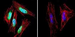
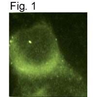
- Immunofluorescent analysis of Presenilin 1 using Presenilin 1 Monoclonal antibody (APS 18) (Product # MA1-752) shows staining in A2058 melanoma cells. Presenilin 1 staining (green), F-Actin staining with Phalloidin (red) and nuclei with DAPI (blue) is shown. Cells were grown on chamber slides and fixed with formaldehyde prior to staining. Cells were probed without (control) or with or an antibody recognizing Presenilin 1 (Product # MA1-752) at a dilution of 1:20-1:100 over night at 4 °C, washed with PBS and incubated with a DyLight-488 conjugated secondary antibody (Product # 35552 for GAR, Product # 35503 for GAM). Images were taken at 60X magnification.
- Submitted by
- Invitrogen Antibodies (provider)
- Main image

- Experimental details
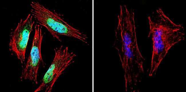
- Immunofluorescent analysis of Presenilin 1 using Presenilin 1 Monoclonal antibody (APS 18) (Product # MA1-752) shows staining in HeLa cells. Presenilin 1 staining (green), F-Actin staining with Phalloidin (red) and nuclei with DAPI (blue) is shown. Cells were grown on chamber slides and fixed with formaldehyde prior to staining. Cells were probed without (control) or with or an antibody recognizing Presenilin 1 (Product # MA1-752) at a dilution of 1:20-1:100 over night at 4 °C, washed with PBS and incubated with a DyLight-488 conjugated secondary antibody (Product # 35552 for GAR, Product # 35503 for GAM). Images were taken at 60X magnification.
- Submitted by
- Invitrogen Antibodies (provider)
- Main image

- Experimental details
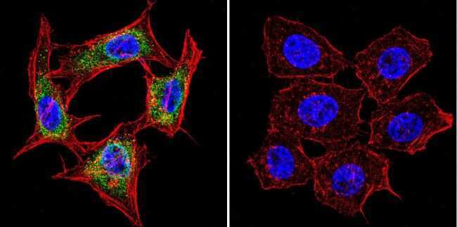
- Immunofluorescent analysis of Presenilin 1 using Presenilin 1 Monoclonal antibody (APS 18) (Product # MA1-752) shows staining in MCF-7 cells. Presenilin 1 staining (green), F-Actin staining with Phalloidin (red) and nuclei with DAPI (blue) is shown. Cells were grown on chamber slides and fixed with formaldehyde prior to staining. Cells were probed without (control) or with or an antibody recognizing Presenilin 1 (Product # MA1-752) at a dilution of 1:20-1:100 over night at 4 °C, washed with PBS and incubated with a DyLight-488 conjugated secondary antibody (Product # 35552 for GAR, Product # 35503 for GAM). Images were taken at 60X magnification.
- Submitted by
- Invitrogen Antibodies (provider)
- Main image

- Experimental details
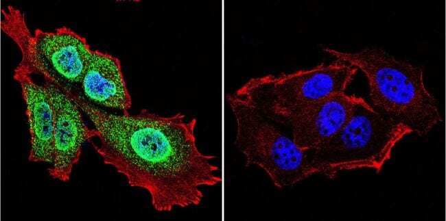
- Immunocytochemical staining of PS1 in mouse fibroblasts using Product # MA1-752.
Supportive validation
- Submitted by
- Invitrogen Antibodies (provider)
- Main image
- Experimental details
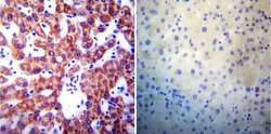
- Immunohistochemistry was performed on normal biopsies of deparaffinized Human liver tissue. To expose target proteins, heat induced antigen retrieval was performed using 10mM sodium citrate (pH6.0) buffer, microwaved for 8-15 minutes. Following antigen retrieval tissues were blocked in 3% BSA-PBS for 30 minutes at room temperature. Tissues were then probed at a dilution of 1:200 with a mouse monoclonal antibody recognizing Presenilin 1 (Product # MA1-752) or without primary antibody (negative control) overnight at 4°C in a humidified chamber. Tissues were washed extensively with PBST and endogenous peroxidase activity was quenched with a peroxidase suppressor. Detection was performed using a biotin-conjugated secondary antibody and SA-HRP, followed by colorimetric detection using DAB. Tissues were counterstained with hematoxylin and prepped for mounting.

- Submitted by
- Invitrogen Antibodies (provider)
- Main image

- Experimental details
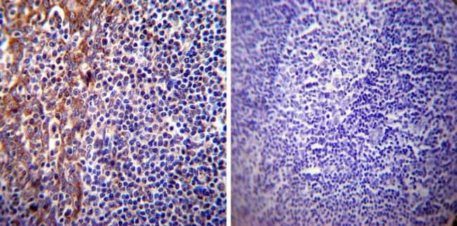
- Immunohistochemistry was performed on normal biopsies of deparaffinized Human tonsil tissue. To expose target proteins, heat induced antigen retrieval was performed using 10mM sodium citrate (pH6.0) buffer, microwaved for 8-15 minutes. Following antigen retrieval tissues were blocked in 3% BSA-PBS for 30 minutes at room temperature. Tissues were then probed at a dilution of 1:20 with a mouse monoclonal antibody recognizing Presenilin 1 (Product # MA1-752) or without primary antibody (negative control) overnight at 4°C in a humidified chamber. Tissues were washed extensively with PBST and endogenous peroxidase activity was quenched with a peroxidase suppressor. Detection was performed using a biotin-conjugated secondary antibody and SA-HRP, followed by colorimetric detection using DAB. Tissues were counterstained with hematoxylin and prepped for mounting.
Supportive validation
- Submitted by
- Invitrogen Antibodies (provider)
- Main image

- Experimental details
- NULL