Explore
 Validate
 Learn
Learn Western blot
Antibody data
- Antibody Data
- Antigen structure
- References [0]
- Comments [0]
- Validations
- Western blot [2]
- Immunocytochemistry [2]
- Immunohistochemistry [3]
Submit
Validation data
Reference
Comment
Report error
- Product number
- MAB7280-100 - Provider product page

- Provider
- R&D Systems
- Product name
- Human/Mouse/Rat CaM Kinase II Pan Specific Antibody
- Antibody type
- Monoclonal
- Description
- Protein A or G purified from hybridoma culture supernatant. Detects human CaM Kinase II gamma in direct ELISAs. Detects human CaM Kinase II alpha , beta , gamma , and delta in Western blots. Detects human, mouse, and rat CaM Kinase II in Western blots.
- Reactivity
- Human, Mouse, Rat
- Host
- Mouse
- Conjugate
- Unconjugated
- Antigen sequence
Q13555- Isotype
- IgG
- Antibody clone number
- 990714
- Vial size
- 100 ug
- Storage
- Use a manual defrost freezer and avoid repeated freeze-thaw cycles. 12 months from date of receipt, -20 to -70 °C as supplied. 1 month, 2 to 8 °C under sterile conditions after reconstitution. 6 months, -20 to -70 °C under sterile conditions after reconstitution.
No comments: Submit comment
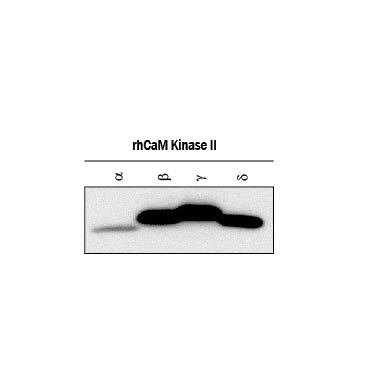
Supportive validation
- Submitted by
- R&D Systems (provider)
- Main image
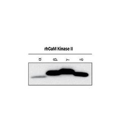
- Experimental details
- Detection of Recombinant Human CaM Kinase II alpha , beta gamma , and delta by Western Blot. Western blot shows lysates of recombinant human CaM Kinase II alpha , recombinant human CaM Kinase II beta , recombinant human CaM Kinase II gamma , and recombinant human CaM Kinase II delta . PVDF membrane was probed with 1 µg/mL of Mouse Anti-Human/Mouse/Rat CaM Kinase II Pan Specific Monoclonal Antibody (Catalog # MAB7280) followed by HRP-conjugated Anti-Sheep IgG Secondary Antibody (Catalog # HAF016). This experiment was conducted under reducing conditions and using Immunoblot Buffer Group 1.
- Submitted by
- R&D Systems (provider)
- Main image
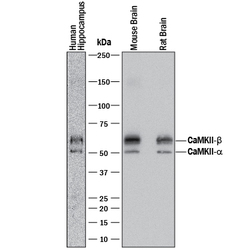
- Experimental details
- Detection of Human, Mouse, and Rat CaM Kinase II by Western Blot. Western blot shows lysates of human brain (hippocampus) tissue, mouse brain tissue, and rat brain tissue. PVDF membrane was probed with 1 µg/mL of Mouse Anti-Human/Mouse/Rat CaM Kinase II Pan Specific Monoclonal Antibody (Catalog # MAB7280) followed by HRP-conjugated Anti-Mouse IgG Secondary Antibody (Catalog # HAF018). A specific band was detected for CaM Kinase II at approximately 50 and 60 kDa (as indicated). This experiment was conducted under reducing conditions and using Immunoblot Buffer Group 1.

Supportive validation
- Submitted by
- R&D Systems (provider)
- Main image
- Experimental details
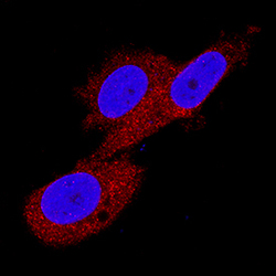
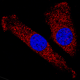
- CaM Kinase II in PC-3 Human Cell Line. CaM Kinase II was detected in immersion fixed PC-3 human prostate cancer cell line using Mouse Anti-Human/Mouse/Rat CaM Kinase II Pan Specific Monoclonal Antibody (Catalog # MAB7280) at 8 µg/mL for 3 hours at room temperature. Cells were stained using the NorthernLights™ 557-conjugated Anti-Mouse IgG Secondary Antibody (red; Catalog # NL007) and counterstained with DAPI (blue). Specific staining was localized to cytoplasm. View our protocol for Fluorescent ICC Staining of Cells on Coverslips.
- Submitted by
- R&D Systems (provider)
- Main image

- Experimental details
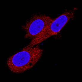
- CaM Kinase II in C2C12 Mouse Cell Line. CaM Kinase II was detected in immersion fixed C2C12 mouse myoblast cell line using Mouse Anti-Human/Mouse/Rat CaM Kinase II Pan Specific Monoclonal Antibody (Catalog # MAB7280) at 25 µg/mL for 3 hours at room temperature. Cells were stained using the NorthernLights™ 557-conjugated Anti-Mouse IgG Secondary Antibody (red; Catalog # NL007) and counterstained with DAPI (blue). Specific staining was localized to cytoplasm. View our protocol for Fluorescent ICC Staining of Cells on Coverslips.
Supportive validation
- Submitted by
- R&D Systems (provider)
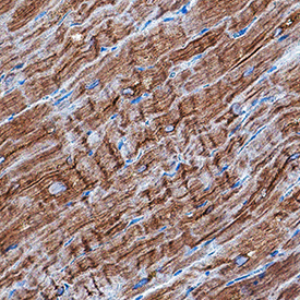
- Main image
- Experimental details
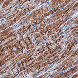
- CaM Kinase II in Human Heart. CaM Kinase II was detected in immersion fixed paraffin-embedded sections of human heart using Mouse Anti-Human/Mouse/Rat CaM Kinase II Pan Specific Monoclonal Antibody (Catalog # MAB7280) at 0.5 µg/mL for 1 hour at room temperature. Tissue was stained using DAB (brown) and counterstained with hematoxylin (blue). Specific staining was localized to cytoplasm in cardiomyocytes. View our protocol for IHC Staining with VisUCyte HRP Polymer Detection Reagents.
- Submitted by
- R&D Systems (provider)
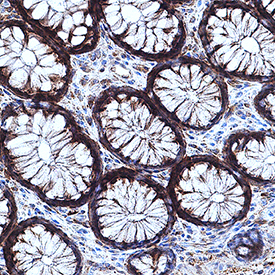
- Main image

- Experimental details
- CaM Kinase II in Human Intestine. CaM Kinase II was detected in immersion fixed paraffin-embedded sections of human intestine using Mouse Anti-Human/Mouse/Rat CaM Kinase II Pan Specific Monoclonal Antibody (Catalog # MAB7280) at 1.7 µg/mL for 1 hour at room temperature. Tissue was stained using DAB (brown) and counterstained with hematoxylin (blue). Specific staining was localized to cytoplasm in intestinal gland cells. View our protocol for IHC Staining with VisUCyte HRP Polymer Detection Reagents.
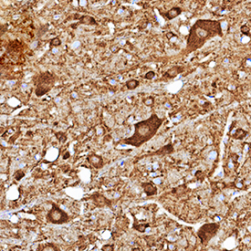
- Submitted by
- R&D Systems (provider)
- Main image

- Experimental details
- CaM Kinase II in Mouse Brain. CaM Kinase II was detected in perfusion fixed frozen sections of mouse brain (brainstem) using Mouse Anti-Human/Mouse/Rat CaM Kinase II Pan Specific Monoclonal Antibody (Catalog # MAB7280) at 1.7 µg/mL for 1 hour at room temperature. Tissue was stained using DAB (brown) and counterstained with hematoxylin (blue). Specific staining was localized to cytoplasm in neurons. View our protocol for IHC Staining with VisUCyte HRP Polymer Detection Reagents.