Explore
 Validate
 Learn
Learn Western blot
 Immunocytochemistry
Antibody data
- Antibody Data
- Antigen structure
- References [1]
- Comments [0]
- Validations
- Immunocytochemistry [2]
- Immunohistochemistry [3]
- Other assay [1]
Submit
Validation data
Reference
Comment
Report error
- Product number
- PA5-96529 - Provider product page

- Provider
- Invitrogen Antibodies
- Product name
- Osteocalcin Polyclonal Antibody
- Antibody type
- Polyclonal
- Antigen
- Synthetic peptide
- Description
- Immunogen sequence: MRALTLLALL ALAALCIAGQ AGAKPSGAES SKGAAFVSKQ EGSEVVKRPR RYLYQWLGAP VPYPDPLEPR REVCELNPDC DELADHIGFQ EAYRRFYGPV
- Reactivity
- Human, Mouse, Rat
- Host
- Rabbit
- Isotype
- IgG
- Vial size
- 100 µL
- Concentration
- 1 mg/mL
- Storage
- -20° C, Avoid Freeze/Thaw Cycles
Submitted references Mechanics-driven nuclear localization of YAP can be reversed by N-cadherin ligation in mesenchymal stem cells.
Zhang C, Zhu H, Ren X, Gao B, Cheng B, Liu S, Sha B, Li Z, Zhang Z, Lv Y, Wang H, Guo H, Lu TJ, Xu F, Genin GM, Lin M
Nature communications 2021 Oct 28;12(1):6229
Nature communications 2021 Oct 28;12(1):6229
No comments: Submit comment
Supportive validation
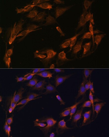
- Submitted by
- Invitrogen Antibodies (provider)
- Main image
- Experimental details
- Immunocytochemistry-Immunofluorescence analysis of Osteocalcin was performed in C6 cells using Osteocalcin Polyclonal Antibody (Product # PA5-96529) at a dilution of 1:100. Blue: DAPI for nuclear staining.
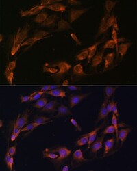
- Submitted by
- Invitrogen Antibodies (provider)
- Main image

- Experimental details
- Immunocytochemistry-Immunofluorescence analysis of Osteocalcin was performed in U-2 OS cells using Osteocalcin Polyclonal Antibody (Product # PA5-96529) at a dilution of 1:100. Blue: DAPI for nuclear staining.
Supportive validation
- Submitted by
- Invitrogen Antibodies (provider)
- Main image
- Experimental details
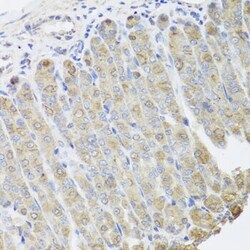
- Immunohistochemistry analysis of Osteocalcin in paraffin-embedded mouse stomach using Osteocalcin Polyclonal Antibody (Product # PA5-96529) at a dilution of 1:100.
- Submitted by
- Invitrogen Antibodies (provider)
- Main image
- Experimental details
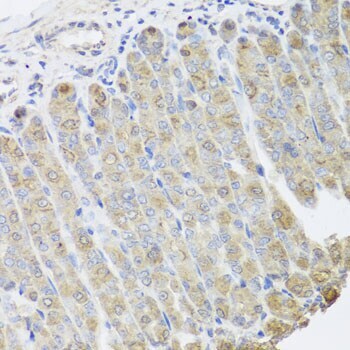
- Immunohistochemistry analysis of Osteocalcin in paraffin-embedded human stomach using Osteocalcin Polyclonal Antibody (Product # PA5-96529) at a dilution of 1:100.
- Submitted by
- Invitrogen Antibodies (provider)
- Main image
- Experimental details
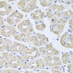
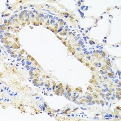
- Immunohistochemistry analysis of Osteocalcin in paraffin-embedded mouse lung using Osteocalcin Polyclonal Antibody (Product # PA5-96529) at a dilution of 1:100.

Supportive validation
- Submitted by
- Invitrogen Antibodies (provider)
- Main image

- Experimental details
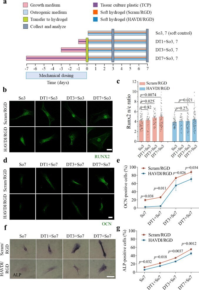
- Fig. 4 The HAVDI/RGD hydrogel can be used to reduce long-term retention of the effects of mechanotransduction and inhibit osteogenic differentiation of hMSCs. a Schematic of the experimental protocol for assessing osteogenic differentiation of hMSCs cultured on TCP for 1-7 d, followed by 3 d (for RUNX2) or 7 d (for OCN and ALP) culture on 20 kPa Scram/RGD or HAVDI/RGD hydrogels. hMSCs were cultured on TCP (dark purple) in growth medium (pink) for 1, 3, or 7 d and then transferred (light yellow, day 0) to soft Scram/RGD (orange) or HAVDI/RGD (blue) hydrogels for additional 3 d or 7 d of culture in osteogenic medium (light blue) before collection and analysis (gray, day 3 or day 7). RUNX2 expression was assessed in the conditions denoted So3 (soft control, without culture on TCP), DT1 + So3, DT3 + So3, or DT7 + So3. OCN and ALP expression were assessed in the conditions denoted So7 (soft control, without culture on TCP), DT1 + So7, DT3 + So7, or DT7 + So7. b Representative confocal images of cells stained for RUNX2, a marker of early osteogenic differentiation, in hMSCs following treatment as shown in a . Scale bars, 20 um. c Analysis of confocal images showed a significant increase in the RUNX2 n/c ratio with increasing exposure to TCP in hMSCs transferred to Scram/RGD hydrogels (from left to right n = 77, 49, 59, 73, 72, 44, 84, 92 cells examined over 7, 15, 19, 20, 12, 24, 23, 26 images respectively, p -values were obtained using one-way ANOVA followed by Tukey's post hoc te