 Explore
 Validate
 Learn
Learn Western blot
Antibody data
- Antibody Data
- Antigen structure
- References [1]
- Comments [0]
- Validations
- Western blot [3]
- ELISA [1]
- Immunocytochemistry [1]
- Immunohistochemistry [3]
Submit
Validation data
Reference
Comment
Report error
- Product number
- GTX101162 - Provider product page

- Provider
- GeneTex
- Proper citation
- GeneTex Cat#GTX101162, RRID:AB_1950920
- Product name
- MIF antibody [N1C3]
- Antibody type
- Polyclonal
- Reactivity
- Human, Mouse, Rat
- Host
- Rabbit
Submitted references Macrophage Migration Inhibitory Factor (MIF) Supports Homing of Osteoclast Precursors to Peripheral Osteolytic Lesions.
Movila A, Ishii T, Albassam A, Wisitrasameewong W, Howait M, Yamaguchi T, Ruiz-Torruella M, Bahammam L, Nishimura K, Van Dyke T, Kawai T
Journal of bone and mineral research : the official journal of the American Society for Bone and Mineral Research 2016 Sep;31(9):1688-700
Journal of bone and mineral research : the official journal of the American Society for Bone and Mineral Research 2016 Sep;31(9):1688-700
No comments: Submit comment
Supportive validation
- Submitted by
- GeneTex (provider)
- Main image

- Experimental details
- Sample (30 ?g of whole cell lysate)A: Molt-4 (GTX27912)15% SDS PAGEGTX101162 diluted at 1:1000The HRP-conjugated anti-rabbit IgG antibody (GTX213110-01) was used to detect the primary antibody.

- Submitted by
- GeneTex (provider)
- Main image

- Experimental details
- Sample (30 ?g of whole cell lysate) A:NIH-3T315% SDS PAGE GTX101162 diluted at 1:1000 The HRP-conjugated anti-rabbit IgG antibody (GTX213110-01) was used to detect the primary antibody.

- Submitted by
- GeneTex (provider)
- Main image

- Experimental details
- Various tissue extracts (50 ?g) were separated by 15% SDS-PAGE, and the membrane was blotted with MIF antibody [N1C3] (GTX101162) diluted at 1:1000. The HRP-conjugated anti-rabbit IgG antibody (GTX213110-01) was used to detect the primary antibody, and the signal was developed with Trident ECL plus-Enhanced.

Supportive validation
- Submitted by
- GeneTex (provider)
- Main image
- Experimental details
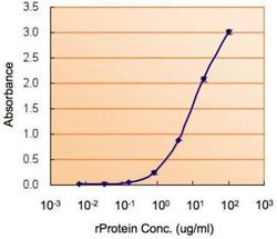
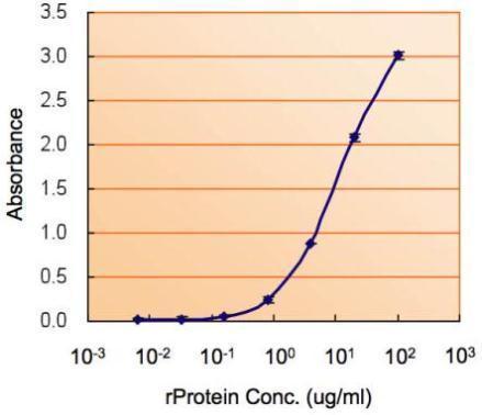
- ELISA detection of MIF using GTX101162 for capture at a concentration of 5 ?g/mL and GTX89535 for detection at a concentration of 1.5 ?g/mL.
Supportive validation
- Submitted by
- GeneTex (provider)
- Main image

- Experimental details
- Immunofluorescence analysis of methanol-fixed HeLa, using MIF(GTX101162) antibody at 1:200 dilution.

Supportive validation
- Submitted by
- GeneTex (provider)
- Main image

- Experimental details
- Immunohistochemical analysis of paraffin-embedded SW480 xenograft, using MIF(GTX101162) antibody at 1:500 dilution.

- Submitted by
- GeneTex (provider)
- Main image

- Experimental details
- MIF antibody [N1C3] detects MIF protein at cytoplasm in mouse brain by immunohistochemical analysis. Sample: Paraffin-embedded mouse brain. MIF antibody [N1C3] (GTX101162) diluted at 1:500.

- Submitted by
- GeneTex (provider)
- Main image

- Experimental details
- MIF antibody [N1C3] detects MIF protein at cytoplasm in rat brain by immunohistochemical analysis. Sample: Paraffin-embedded rat brain. MIF antibody [N1C3] (GTX101162) diluted at 1:500.
