 Explore
 Validate
 Learn
Learn Western blot
 Immunohistochemistry
Antibody data
- Antibody Data
- Antigen structure
- References [0]
- Comments [0]
- Validations
- Western blot [1]
- Immunohistochemistry [6]
Submit
Validation data
Reference
Comment
Report error
- Product number
- AMAb91561 - Provider product page

- Provider
- Atlas Antibodies
- Product name
- Anti-AMACR
- Antibody type
- Monoclonal
- Reactivity
- Human
- Host
- Mouse
- Conjugate
- Unconjugated
- Antigen sequence
APFYTTYRTADGEFMAVGAIEPQFYELLIKGLGLK
SDELPNQMSMDDWPEMKKKFADVFAKKTKAEWCQI
FDGTDACVTPVLTF- Epitope
- Binds to an epitope located within the peptide sequence EPQFYELLIK as determined by overlapping synthetic peptides.
- Isotype
- IgG
- Antibody clone number
- CL9360
- Vial size
- 100 µl
- Storage
- Store at +4°C for short term storage. Long time storage is recommended at -20°C.
No comments: Submit comment
Supportive validation
- Submitted by
- Atlas Antibodies (provider)
- Main image

- Experimental details
- Western blot analysis in human cell line CACO-2.

Enhanced validation
Supportive validation
- Submitted by
- Atlas Antibodies (provider)
- Enhanced method
- Orthogonal validation
- Main image
- Experimental details
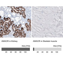
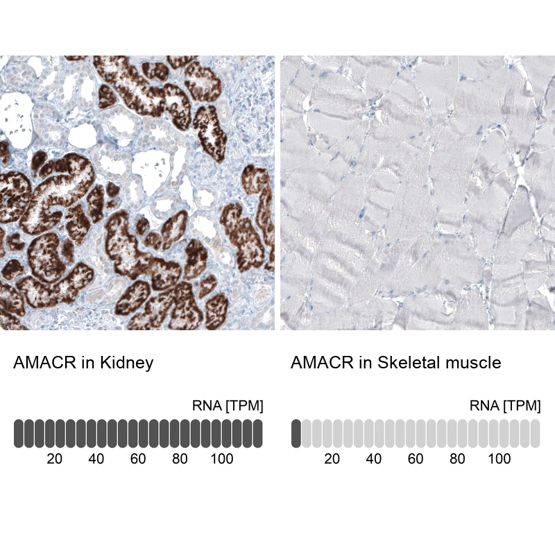
- Immunohistochemistry analysis in human kidney and skeletal muscle tissues using AMAb91561 antibody. Corresponding AMACR RNA-seq data are presented for the same tissues.
- Sample type
- HUMAN
Supportive validation
- Submitted by
- Atlas Antibodies (provider)
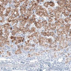
- Main image

- Experimental details
- Immunohistochemical staining of human pancreas shows weak granular cytoplasmic positivity in islets of Langerhans.

- Submitted by
- Atlas Antibodies (provider)
- Main image
- Experimental details
- Immunohistochemical staining of human liver cancer shows moderate granular cytoplasmic positivity in tumor cells.

- Submitted by
- Atlas Antibodies (provider)
- Main image

- Experimental details
- Immunohistochemical staining of human liver shows moderate granular cytoplasmic positivity in hepatocytes.

- Submitted by
- Atlas Antibodies (provider)
- Main image

- Experimental details
- Immunohistochemical staining of human skeletal muscle shows no positivity in myocytes as expected.

- Submitted by
- Atlas Antibodies (provider)
- Main image

- Experimental details
- Immunohistochemical staining of human kidney shows strong granular cytoplasmic positivity in cells in proximal tubules.
