 Explore
 Validate
 Learn
Learn Immunocytochemistry
Antibody data
- Antibody Data
- Antigen structure
- References [3]
- Comments [0]
- Validations
- Immunocytochemistry [4]
- Immunohistochemistry [1]
- Flow cytometry [2]
- Other assay [1]
Submit
Validation data
Reference
Comment
Report error
- Product number
- MA1-022 - Provider product page

- Provider
- Invitrogen Antibodies
- Product name
- SSEA1 Monoclonal Antibody (MC-480)
- Antibody type
- Monoclonal
- Antigen
- Other
- Reactivity
- Human, Mouse
- Host
- Mouse
- Isotype
- IgM
- Antibody clone number
- MC-480
- Vial size
- 100 µg
- Concentration
- 1 mg/mL
- Storage
- -20°C
Submitted references Comparison of in vitro Neuronal Differentiation Capacity Between Mouse Epiblast Stem Cells Derived From Nuclear Transfer and Naturally Fertilized Embryos.
USP26 functions as a negative regulator of cellular reprogramming by stabilising PRC1 complex components.
Amnion Epithelial Cells of Buffalo (Bubalus Bubalis) Term Placenta Expressed Embryonic Stem Cells Markers and Differentiated into Cells of Neurogenic Lineage In Vitro.
Li T, Zheng Y, Li Y, Ye D
Frontiers in molecular neuroscience 2018;11:392
Frontiers in molecular neuroscience 2018;11:392
USP26 functions as a negative regulator of cellular reprogramming by stabilising PRC1 complex components.
Ning B, Zhao W, Qian C, Liu P, Li Q, Li W, Wang RF
Nature communications 2017 Aug 24;8(1):349
Nature communications 2017 Aug 24;8(1):349
Amnion Epithelial Cells of Buffalo (Bubalus Bubalis) Term Placenta Expressed Embryonic Stem Cells Markers and Differentiated into Cells of Neurogenic Lineage In Vitro.
Ghosh K, Selokar NL, Gahlawat SK, Kumar D, Kumar P, Yadav PS
Animal biotechnology 2016;27(1):38-43
Animal biotechnology 2016;27(1):38-43
No comments: Submit comment
Supportive validation
- Submitted by
- Invitrogen Antibodies (provider)
- Main image

- Experimental details
- Immunofluorescent analysis of SSEA-1 using anti-SSEA-1 monoclonal antibody (Product # MA1-022) shows staining on the cell surface of human differentiated HEL 11.4 iPS cells, indicating loss of pluripotency. SSEA-1 staining (green) and an overlay image of SSEA-1 with DAPI (blue) is shown. HEL 11.4 cells were grown on matrigel coated chamber slides and fixed with formaldehyde prior to staining. Cells were probed with a mouse monoclonal antibody recognizing SSEA-1 (Product # MA1-022) at a dilution of 1:120 over night at 4°C, washed with PBS and incubated with a FITC-conjugated secondary antibody at a dilution of 1:100 for 60 minutes at room temperature. Images were taken at 20X magnification.

- Submitted by
- Invitrogen Antibodies (provider)
- Main image

- Experimental details
- Immunofluorescent analysis of SSEA-1 using anti-SSEA-1 monoclonal antibody (Product # MA1-022) shows staining on the cell surface of peripheral human embryonic H9 stem cells, indicating partial loss of pluripotency. SSEA-1 staining (green) and an overlay image of SSEA-1 with DAPI (blue) is shown. H9 cells were grown on matrigel coated chamber slides and fixed with formaldehyde prior to staining. Cells were probed with a mouse monoclonal antibody recognizing SSEA-1 (Product # MA1-022) at a dilution of 1:120 over night at 4°C, washed with PBS and incubated with a FITC-conjugated secondary antibody at a dilution of 1:100 for 60 minutes at room temperature. Images were taken at 20X magnification.

- Submitted by
- Invitrogen Antibodies (provider)
- Main image

- Experimental details
- Immunofluorescent analysis of SSEA1 (red) in mouse embryonic CJ7 stem cells grown on 0.1% gelatin. The cells were fixed with 4% paraformaldehyde at room temperature for 10 min and permeabilized with 0.25% Triton-X 100 for 5 min and blocked with 10% BSA in PBS for 30 min at 37°C. Cells were stained with a SSEA1 monoclonal antibody (Product # MA1-022) at a dilution of 1:200 in 3% BSA/PBS blocking buffer overnight at 4°C and then incubated with a RRX-conjugated donkey anti-mouse IgG secondary antibody at a dilution of 1:500 for 1 hour at room temperature. Nucleus DNA (blue) was stained with DAPI (Product # D1306). Note: Data courtesy of Innovators program.

- Submitted by
- Invitrogen Antibodies (provider)
- Main image

- Experimental details
- Immunofluorescent analysis of SSEA-1 using anti-SSEA-1 monoclonal antibody (Product # MA1-022) shows specific expression in mouse testicular teratoma F9 cells (shown in green) but not in negative control human NTERA-2 teratocarcinoma cells. Formalin fixed cells were permeabilized with 0.1% Triton X-100 in TBS for 10 minutes at room temperature. Cells were blocked with 1% Blocker BSA (Product # 37525) for 15 minutes at room temperature. Cells were probed with a mouse monoclonal antibody recognizing SSEA-1 (Product # MA1-022), at a dilution of 1:50 for at least 1 hour at room temperature. Cells were washed with PBS and incubated with a fluorescent dye labeled goat-anti-mouse IgM secondary antibody at a dilution of 1:400 for 30 minutes at room temperature. Nuclei (blue) were stained with Hoechst 33342 dye (Product # 62249). Images were taken on a Thermo Scientific ArrayScan at 20X magnification.

Supportive validation
- Submitted by
- Invitrogen Antibodies (provider)
- Main image

- Experimental details
- Immunohistochemistry was performed on frozen sections of Knifefish (Apteronotus leptorhynchus) brain tissue fixed with 2% paraformaldehyde and permeabilized with 0.3% Triton X-100. Tissue sections were washed with TBS and blocked in a solution containing 3% serum, 1% BSA, 1% teleostean gelatin, 0.3% Triton X-100 in TBS, for 1 hour at room temperature. The primary antibody SSEA1 (Product # MA1-022X) (1:50 dilution) was applied overnight (~18 hours) at 4°C. After washing with TBS, an Alexa Fluor® 546 - conjugated goat anti-mouse secondary antibody was applied for 2 hours at room temperature at a 1:200 dilution. Sections were counterstained with DAPI. Images were taken with a Zeiss 710 confocal microscope at 63X magnification. The antibody shows specific surface staining of blood vessels in the brain, shown here within a proliferation region in the optic tectum in the brain. In addition, the antibody labeled the surface of certain myelinated axons and did not stain Knifefish stem/progenitor cells. Calibration bar = 10µm. Note: Data courtesy of Innovators program.

Supportive validation
- Submitted by
- Invitrogen Antibodies (provider)
- Main image

- Experimental details
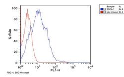
- Flow cytometry analysis of SSEA-1 (blue histogram) on H9 human embryonic stem cells. Cells were harvested, fixed with 4% formaldehyde, washed with PBS, and incubated with a SSEA-1 monoclonal antibody (Product # MA1-022) or an IgM isotype control (red histogram) at a 1:100 dilution for 1 hour on ice. For flow analysis, a 30-minute incubation with FITC-conjugated secondary antibody was performed and 100,000 cells were acquired for each sample.

- Submitted by
- Invitrogen Antibodies (provider)
- Main image
- Experimental details
- Flow cytometry analysis of SSEA-1 (blue histogram) on differentiated HEL 11.4 human iPS cells. Cells were harvested, fixed with 4% formaldehyde, washed with PBS, and incubated with a SSEA-1 monoclonal antibody (Product # MA1-022) or an IgM isotype control (red histogram) at a 1:100 dilution for 1 hour on ice. For flow analysis, a 30-minute incubation with FITC-conjugated secondary antibody was performed and 100,000 cells were acquired for each sample.

Supportive validation
- Submitted by
- Invitrogen Antibodies (provider)
- Main image

- Experimental details
- FIGURE 1 Derivation and characterization of mouse EpiSCs from in vivo fertilization and NT embryos. (A) Mouse embryo at E5.5 stage, isolated epiblast layer, and morphology of established F-mEpiSCs and NT-mEpiSCs. (B) Immunofluorescence staining for Nanog, SSEA1, and Oct4 in F-EpiSCs and NT-EpiSCs; nuclei are shown using DAPI. (C) Gene expression analysis by qPCR for EpiSC-specific markers, pluripotency markers, and neural marker in four selected mEpiSCs. Scale bar: 100 mum; n.s., P > 0.05; ** P < 0.01.
