Explore
 Validate
 Learn
Learn Western blot
 ELISA
ELISAAntibody data
- Antibody Data
- Antigen structure
- References [2]
- Comments [0]
- Validations
- Western blot [1]
- Immunocytochemistry [1]
- Immunohistochemistry [1]
- Other assay [2]
Submit
Validation data
Reference
Comment
Report error
- Product number
- MA5-15331 - Provider product page

- Provider
- Invitrogen Antibodies
- Product name
- CD34 Monoclonal Antibody (4H5E7, 9B10D4)
- Antibody type
- Monoclonal
- Antigen
- Purifed from natural sources
- Description
- Applications Tested: This C8.6 antibody has been pre-titrated and tested by intracellular staining and flow cytometric analysis of stimulated normal human peripheral blood cells. This can be used at 5 µL (0.25 µg) per test. A test is defined as the amount (µg) of antibody that will stain a cell sample in a final volume of 100 µL. Cell number should be determined empirically but can range from 10^5 to 10^8 cells/test.
- Reactivity
- Human
- Host
- Mouse
- Isotype
- IgG
- Antibody clone number
- 4H5E7, 9B10D4
- Vial size
- 100 µL
- Concentration
- Conc. Not Determined
- Storage
- Store at 4°C short term. For long term storage, store at -20°C, avoiding freeze/thaw cycles.
Submitted references Pathology of Fibrosis in Crohn's Disease-Contribution to Understanding Its Pathogenesis.
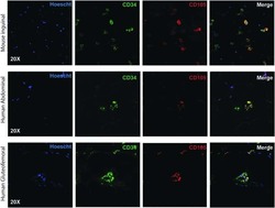
Utilizing Confocal Microscopy to Characterize Human and Mouse Adipose Tissue.
Zidar N, Langner C, Jerala M, Boštjančič E, Drobne D, Tomažič A
Frontiers in medicine 2020;7:167
Frontiers in medicine 2020;7:167
Utilizing Confocal Microscopy to Characterize Human and Mouse Adipose Tissue.
Blackshear CP, Borrelli MR, Shen EZ, Ransom RC, Chung NN, Vistnes SM, Irizarry D, Nazerali R, Momeni A, Longaker MT, Wan DC
Tissue engineering. Part C, Methods 2018 Oct;24(10):566-577
Tissue engineering. Part C, Methods 2018 Oct;24(10):566-577
No comments: Submit comment
Supportive validation
- Submitted by
- Invitrogen Antibodies (provider)
- Main image

- Experimental details
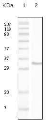
- Western blot analysis of CD34/gp105-120 using a CD34/gp105-120 monoclonal antibody (Product # MA5-15331) against a truncated CD34 recombinant protein.
Supportive validation
- Submitted by
- Invitrogen Antibodies (provider)
- Main image

- Experimental details
- Immunofluorescence analysis of peripheral blood cells using CD34/gp105-120 monoclonal antibody (Product # MA5-15331).

Supportive validation
- Submitted by
- Invitrogen Antibodies (provider)
- Main image

- Experimental details
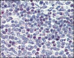
- Immunohistochemical analysis of paraffin-embedded human spleen using CD34/gp105-120 monoclonal antibody (Product # MA5-15331) followed with DAB staining.
Supportive validation
- Submitted by
- Invitrogen Antibodies (provider)
- Main image
- Experimental details
- NULL

- Submitted by
- Invitrogen Antibodies (provider)
- Main image

- Experimental details
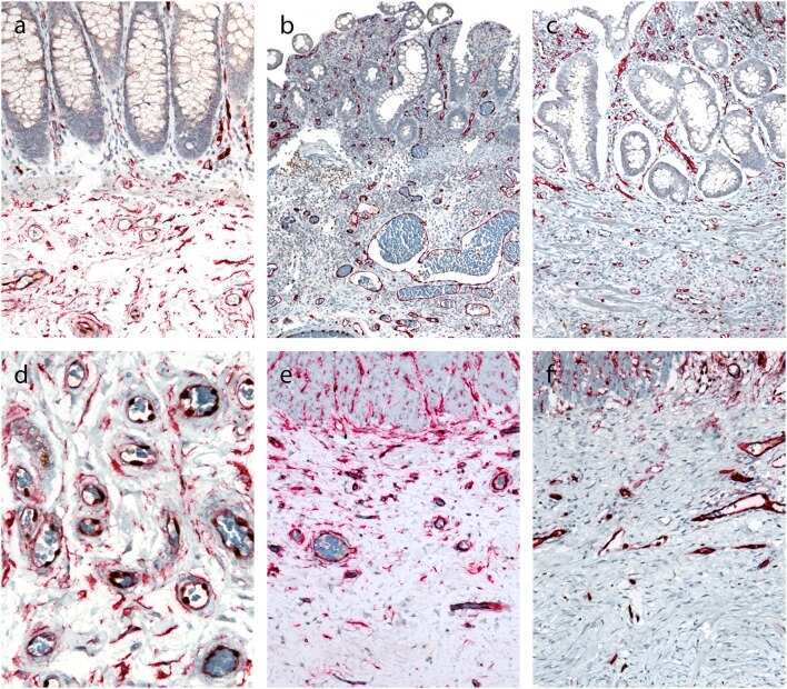
- Figure 4 Double immunohistochemical reaction for CD34 and erg in the normal colon and Crohn's disease with and without stenosis. (a,d) Normal colon: double immunostaining for CD34 (red) and erg (brown) in endothelial cells in blood vessels, and red cytoplasmic reaction in spindle-shaped cells in the submucosa (a) and subserosa, particularly around blood vessels (d) . (b,e) Crohn's disease without stenosis: double immunostaining for CD34 (red) an erg (brown) in endothelial cells in blood vessels in the lamina propria and submucosa; no spindle-shaped cells with red cytoplasmic reaction (b) . Double immunostaining for CD34 (red) and erg (brown) in endothelial cells in blood vessels, and red cytoplasmic reaction in spindle-shaped cells in the subserosa, particularly around blood vessels (e) . (c,f) Crohn's disease with stenosis: double immunostaining for CD34 (red) an erg (brown) in endothelial cells in blood vessels in the lamina propria and submucosa (C) and in fibrosis in subserosa (f) ; no spindle-shaped cells with red cytoplasmic reaction.