Explore
 Validate
 Learn
Learn Western blot
 Immunocytochemistry
Antibody data
- Antibody Data
- Antigen structure
- References [1]
- Comments [0]
- Validations
- Immunocytochemistry [1]
- Flow cytometry [2]
- Other assay [1]
Submit
Validation data
Reference
Comment
Report error
- Product number
- DM1204 - Provider product page

- Provider
- OriGene
- Product name
- CEACAM5 mouse monoclonal antibody, clone D14HD11, Purified
- Antibody type
- Monoclonal
- Description
- CEACAM5 mouse monoclonal antibody, clone D14HD11, Purified
- Host
- Mouse
- Conjugate
- Unconjugated
- Epitope
- CEACAM5
- Isotype
- IgG
- Antibody clone number
- D14HD11
- Vial size
- 100 µg
- Concentration
- 2.0 mg/ml
Submitted references Carcinoembryonic antigen family members CEACAM6 and CEACAM7 are differentially expressed in normal tissues and oppositely deregulated in hyperplastic colorectal polyps and early adenomas.
Schölzel S, Zimmermann W, Schwarzkopf G, Grunert F, Rogaczewski B, Thompson J
The American journal of pathology 2000 Feb;156(2):595-605
The American journal of pathology 2000 Feb;156(2):595-605
No comments: Submit comment
Supportive validation
- Submitted by
- OriGene (provider)
- Main image

- Experimental details
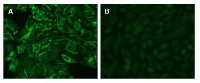
- Figure 2: Immunofluorescence Microscopy of CHO cells using D14HD11 . CHO cells were transfected with an expression vector encoding CEACAM1 (A). Untransfected CHO parental cells served as Negative Control (B). Binding of D14HD11 was visualized with a FITC-conjugated secondary antibody.
- Validation comment
- IF
Supportive validation
- Submitted by
- OriGene (provider)
- Main image

- Experimental details
- Figure 4: FACS analysis of BOSC23 cells using D14HD11 . BOSC23 cells were transiently transfected with anexpression vector encoding either CEACAM1,3,4,5,6 (red curves) or an irrelevant protein (control transfectant: blackcurves). Binding of D14HD11 was detected with a PE-conjugated secondary antibody. A positive signal wasobtained only with CEACAM1, CEACAM3, CEACAM4,CEACAM5 and CEACAM6 expressing cells.
- Validation comment
- FC

- Submitted by
- OriGene (provider)
- Main image

- Experimental details
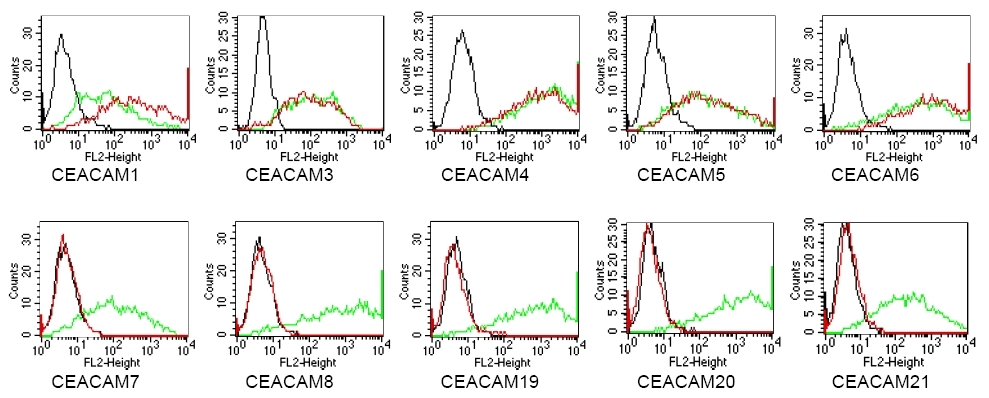
- Figure 3: BOSC cells were transiently transfected with expression vectors containing either the cDNA of CEACAM1, CEACAM3-CEACAM8 or CEACAM19-21. Recognition of CEACAM4 was tested on CHO cells stably transfected with a CEACAM4 expressionvector. Expression of the constructs was confirmed with monoclonal antibodies known to recognise the corresponding proteins(CEACAM1: 4/3/17, CEACAM3,4: D14HD11, CEACAM5: 26/3/13, CEACAM6: 9A6, CEACAM7: BAC2, CEACAM8: GM-2H6,CEACAM19-21: anti-myc, green curves). An irrelevant monoclonal antibody served as a negative control (black curves). Forspecificity testing, protein G purified D14HD11 was tested on all CEACAM transfectants. A positive signal was obtained with CEA-CAM1, CEACAM3, CEACAM4, CEACAM5 and CEACAM6 expressing cells (red curves).
- Validation comment
- FC
Supportive validation
- Submitted by
- OriGene (provider)
- Main image
- Experimental details
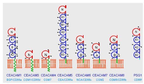
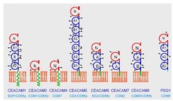
- Figure 1: Domain organization of the human CEACAM family. The CEACAM family consists of two subgroups, the CEACAM and the PSG subgroup. CEACAM family members are membrane bound by either via a transmembrane domain or a GPI anchor (green arrow) whereas the PSGs are secreted glycoproteins. N-linked glycosylation sites are shown as drum sticks. The IgV domains are shown. in red, the IgC domains in blue.
- Validation comment
- Assay