 Explore
 Validate
 Learn
Learn Western blot
 ELISA
ELISAAntibody data
- Antibody Data
- Antigen structure
- References [2]
- Comments [0]
- Validations
- Western blot [6]
- Immunohistochemistry [2]
- Flow cytometry [2]
Submit
Validation data
Reference
Comment
Report error
- Product number
- NBP2-33184 - Provider product page

- Provider
- Novus Biologicals
- Product name
- Mouse Monoclonal GFAP Antibody
- Antibody type
- Monoclonal
- Description
- Protein G purified. This MAb recognizes a protein of ~50kDa which is identified as Glial Fibrillary Acidic Protein (GFAP). It shows no cross-reaction with other intermediate filament proteins. GFAP is specifically found in astroglia. GFAP is a very popular marker for localizing benign astrocyte and neoplastic cells of glial origin in the central nervous system. Antibody to GFAP is useful in differentiating primary gliomas from metastatic lesions in the brain and for documenting astrocytic differentiation in tumors outside the CNS.
- Reactivity
- Human, Mouse, Rat, Bovine, Chicken/Avian, Porcine, Rabbit
- Host
- Mouse
- Isotype
- IgG
- Vial size
- 0.1 mg
- Concentration
- 1.0 mg/ml
- Storage
- Store at 4C short term. Aliquot and store at -20C long term. Avoid freeze-thaw cycles.
Submitted references Transfer and Integration of Breast Milk Stem Cells to the Brain of Suckling Pups.
Nanoparticle fullerol alleviates radiculopathy via NLRP3 inflammasome and neuropeptides.
Aydın MŞ, Yiğit EN, Vatandaşlar E, Erdoğan E, Öztürk G
Scientific reports 2018 Sep 24;8(1):14289
Scientific reports 2018 Sep 24;8(1):14289
Nanoparticle fullerol alleviates radiculopathy via NLRP3 inflammasome and neuropeptides.
Jin L, Ding M, Oklopcic A, Aghdasi B, Xiao L, Li Z, Jevtovic-Todorovic V, Li X
Nanomedicine : nanotechnology, biology, and medicine 2017 Aug;13(6):2049-2059
Nanomedicine : nanotechnology, biology, and medicine 2017 Aug;13(6):2049-2059
No comments: Submit comment
Supportive validation
- Submitted by
- Novus Biologicals (provider)
- Main image
- Experimental details
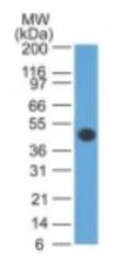
- Western Blot: GFAP Antibody (GA5) - Azide and BSA Free [NBP2-33184] - Analysis showing relative size.

- Submitted by
- Novus Biologicals (provider)
- Main image

- Experimental details
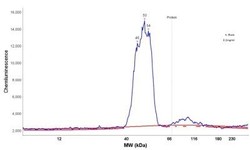
- Simple Western: GFAP Antibody (GA-5) - Azide and BSA Free [NBP2-33184] - Simple Western lane view shows a specific band for GFAP in 0.2 mg/ml of h. Brain lysate(s). This experiment was performed under reducing conditions using the 12-230 kDa separation system.

- Submitted by
- Novus Biologicals (provider)
- Main image
- Experimental details
- Simple Western: GFAP Antibody (GA-5) - Azide and BSA Free [NBP2-33184] - Electropherogram image of the corresponding Simple Western lane. GFAP antibody was used at 10 ug/ml dilution of h. Brain lysates(s) respectively.

- Submitted by
- Novus Biologicals (provider)
- Main image

- Experimental details
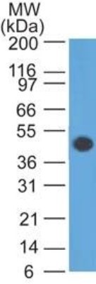
- Western Blot: GFAP Antibody (GA-5) - Azide and BSA Free [NBP2-33184] - Analysis of GFAP in human brain lysate using GFAP (GA5) antibody at 1 ug/ml. goat anti-mouse Ig HRP secondary antibody and PicoTect ECL substrate solution were used for this test.
- Submitted by
- Novus Biologicals (provider)
- Main image

- Experimental details
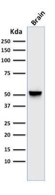
- Western Blot: GFAP Antibody (GA-5) - Azide and BSA Free [NBP2-33184] - Western Blot Analysis of human brain tissue lysate using GFAP Antibody (GA-5).
- Submitted by
- Novus Biologicals (provider)
- Main image

- Experimental details
- Western Blot: GFAP Antibody (GA-5) - Azide and BSA Free [NBP2-33184] - Analysis showing relative size.

Supportive validation
- Submitted by
- Novus Biologicals (provider)
- Main image
- Experimental details
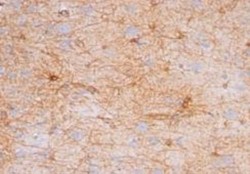
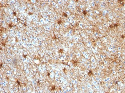
- Immunohistochemistry-Paraffin: GFAP Antibody (GA-5) - Azide and BSA Free [NBP2-33184] - Analysis in human Cerebellum.

- Submitted by
- Novus Biologicals (provider)
- Main image

- Experimental details
- Immunohistochemistry-Paraffin: GFAP Antibody (GA-5) - Azide and BSA Free [NBP2-33184] - Formalin-fixed, paraffin-embedded human Cerebellum stained with GFAP Antibody (GA-5).
Supportive validation
- Submitted by
- Novus Biologicals (provider)
- Main image
- Experimental details
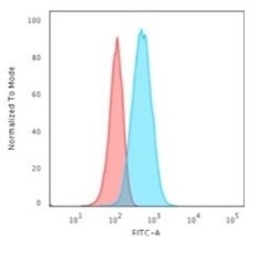
- Flow Cytometry: GFAP Antibody (GA-5) - Azide and BSA Free [NBP2-33184] - Flow Cytometric Analysis of T98G cells using GFAP Antibody (GA-5) followed by Goat anti-Mouse IgG-CF488 (Blue); Isotype Control (Red).

- Submitted by
- Novus Biologicals (provider)
- Main image
- Experimental details
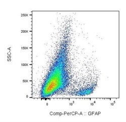
- Flow Cytometry: GFAP Antibody (GA-5) - Azide and BSA Free [NBP2-33184] - Experimental autoimmune encephalomyelitis was induced in C57BL6/J mice, and mononuclear cells were isolated from the CNS at day 10 (onset of symptoms). Cells were stained for GFAP, Neun, CX3CL1, CXCL12, CCL2, CD45 and CD11b, plus for viability to exclude dead cells. GFAP staining is shown for viable cells. Image from verified customer review.
