Explore
 Validate
 Learn
Learn Western blot
Antibody data
- Antibody Data
- Antigen structure
- References [1]
- Comments [0]
- Validations
- Western blot [5]
- Immunocytochemistry [2]
- Immunohistochemistry [9]
Submit
Validation data
Reference
Comment
Report error
- Product number
- GTX100850 - Provider product page

- Provider
- GeneTex
- Proper citation
- GeneTex Cat#GTX100850, RRID:AB_10632188
- Product name
- GFAP antibody
- Antibody type
- Polyclonal
- Reactivity
- Human, Mouse, Rat
- Host
- Rabbit
Submitted references Normalization of T2 relaxation time and apparent diffusion coefficient in relation to the inflammatory changes in the substantia nigra of rats with focal cerebral ischemia.
Yang YM, Li CC, Yin le K, Feng X
Acta radiologica (Stockholm, Sweden : 1987) 2015 Jul;56(7):837-43
Acta radiologica (Stockholm, Sweden : 1987) 2015 Jul;56(7):837-43
No comments: Submit comment
Supportive validation
- Submitted by
- GeneTex (provider)
- Main image

- Experimental details
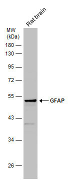
- Sample (50 ug of whole cell lysate) A: Mouse brain 10% SDS PAGE GTX100850 diluted at 1:10000
- Validation comment
- WB
- Submitted by
- GeneTex (provider)
- Main image
- Experimental details
- Sample (30 ug of whole cell lysate) A:U87-MG 10% SDS PAGE GTX100850 diluted at 1:1000
- Validation comment
- WB

- Submitted by
- GeneTex (provider)
- Main image
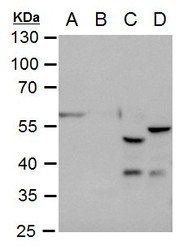
- Experimental details
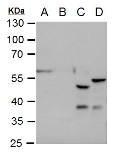
- GFAP antibody detects GFAP protein by western blot analysis.A. 30 £gg U87-MG whole cell extractB. 30 £gg IMR32 whole cell extractC. 50 £gg mouse brain extractD. 50 £gg rat brain extract10 % SDS-PAGEGFAP antibody (GTX100850) dilution: 1:2500
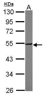
- Submitted by
- GeneTex (provider)
- Main image
- Experimental details
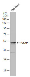
- Rat tissue extract (50 ?g) was separated by 10% SDS-PAGE, and the membrane was blotted with GFAP antibody (GTX100850) diluted at 1:10000.
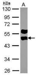
- Submitted by
- GeneTex (provider)
- Main image

- Experimental details
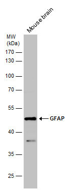
- Mouse tissue extract (50 ?g) was separated by 10% SDS-PAGE, and the membrane was blotted with GFAP antibody (GTX100850) diluted at 1:2500.
Supportive validation
- Submitted by
- GeneTex (provider)
- Main image

- Experimental details
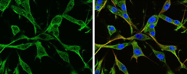
- GFAP antibody detects GFAP protein at cytoplasm by immunofluorescent analysis.Sample: U-87 MG cells were fixed in 4% paraformaldehyde at RT for 15 min.Green: GFAP protein stained by GFAP antibody (GTX100850) diluted at 1:250.Red: beta Tubulin 3/ TUJ1 protein stained by beta Tubulin 3/ TUJ1 antibody (GTX631836) diluted at 1:200.Blue: Hoechst 33342 staining.
- Submitted by
- GeneTex (provider)
- Main image
- Experimental details
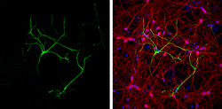
- GFAP antibody detects GFAP protein at glia cells by immunofluorescent analysis.Sample: DIV9 rat E18 primary cortical neurons were fixed in 4% paraformaldehyde at RT for 15 min.Green: GFAP protein stained by GFAP antibody (GTX100850) diluted at 1:500.Red: beta Tubulin 3/ Tuj1, a neuron cell marker, stained by beta Tubulin 3/ Tuj1 antibody [GT11710] (GTX631836) diluted at 1:500.Blue: Fluoroshield with DAPI (GTX30920).
Supportive validation
- Submitted by
- GeneTex (provider)
- Main image
- Experimental details
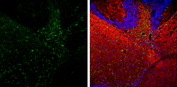
- GFAP antibody detects GFAP protein at astrocyte on mouse fore brain by immunohistochemical analysis. Sample: Paraffin-embedded mouse fore brain. GFAP antibody (GTX100850) diluted at 1:500.

- Submitted by
- GeneTex (provider)
- Main image

- Experimental details
- GFAP antibody detects GFAP protein on embryonic mouse brain by immunohistochemical analysis. Sample:Frozen section of embryonic mouse brain (mE18.5). Green: GFAP antibody (GTX100850) diluted at 1:500. Blue: DAPI

- Submitted by
- GeneTex (provider)
- Main image

- Experimental details
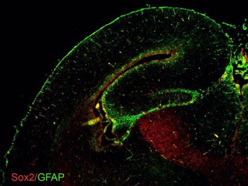
- GFAP antibodies detects GFAP proteins on embryonic mouse brain by immunohistochemical analysis. Sample: Frozen section of embryonic mouse brain (mE18.5). Green: GFAP antibody (GTX100850) diluted at 1:500. Red: Sox2 antibody [GT1876] (GTX627404) diluted at 1:500.
- Submitted by
- GeneTex (provider)
- Main image

- Experimental details
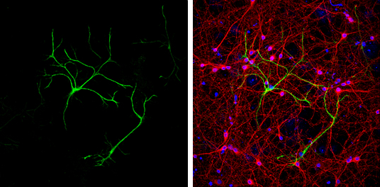
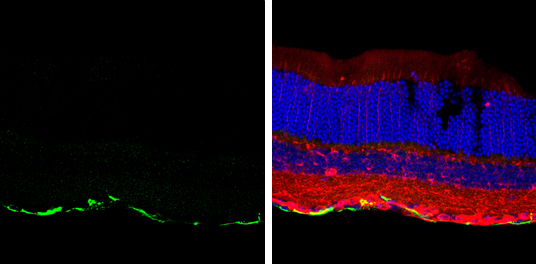
- GFAP antibody detects GFAP protein at retinal ganglion cell layer by immunohistochemical analysis.Sample: Frozen sectioned adult mouse retina. Green: GFAP protein stained by GFAP antibody (GTX100850) diluted at 1:250.Red: beta Tubulin 3/ TUJ1, stained by beta Tubulin 3/ TUJ1 antibody [GT11710] (GTX631836) diluted at 1:250.Blue: Fluoroshield with DAPI (GTX30920).
- Submitted by
- GeneTex (provider)
- Main image
- Experimental details
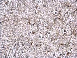
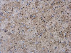
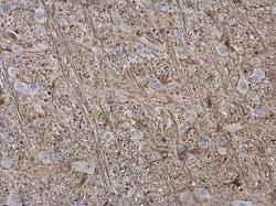
- GFAP antibody detects GFAP protein expression in astrocytes/glia cells on mouse brain by immunohistochemical analysis. Sample: Paraffin-embedded mouse brain. GFAP antibody (GTX100850) diluted at 1:500.
- Submitted by
- GeneTex (provider)
- Main image
- Experimental details
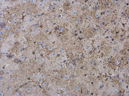
- GFAP antibody detects GFAP protein expression in astrocytes/glia cells on rat brain by immunohistochemical analysis. Sample: Paraffin-embedded rat brain. GFAP antibody (GTX100850) diluted at 1:500.

- Submitted by
- GeneTex (provider)
- Main image
- Experimental details
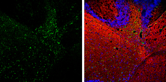
- GFAP antibody detects GFAP protein expression by immunohistochemical analysis.Sample: Frozen-sectioned adult mouse cerebellum. Green: GFAP protein stained by GFAP antibody (GTX100850) diluted at 1:250.Red: beta Tubulin 3/ TUJ1, stained by beta Tubulin 3/ TUJ1 antibody [GT11710] (GTX631836) diluted at 1:500.Blue: Fluoroshield with DAPI (GTX30920).
- Submitted by
- GeneTex (provider)
- Main image

- Experimental details
- GFAP antibody detects GFAP protein expression by immunohistochemical analysis.Sample: Frozen-sectioned adult mouse cerebellum. Green: GFAP protein stained by GFAP antibody (GTX100850) diluted at 1:250.Red: beta Tubulin 3/ TUJ1, stained by beta Tubulin 3/ TUJ1 antibody [GT11710] (GTX631836) diluted at 1:500.Blue: Fluoroshield with DAPI (GTX30920).
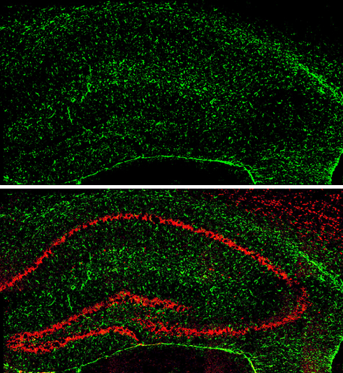
- Submitted by
- GeneTex (provider)
- Main image

- Experimental details
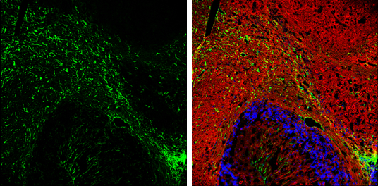
- GFAP antibody detects GFAP protein expression by immunohistochemical analysis.Sample: Frozen-sectioned adult mouse hippocampus. Green: GFAP protein stained by GFAP antibody (GTX100850) diluted at 1:250.Red: NeuN, stained by NeuN antibody [2Q158] (GTX30773) diluted at 1:500.