Antibody data
- Antibody Data
- Antigen structure
- References [8]
- Comments [0]
- Validations
- Western blot [6]
- Immunocytochemistry [1]
- Immunohistochemistry [6]
Submit
Validation data
Reference
Comment
Report error
- Product number
- GTX108711 - Provider product page

- Provider
- GeneTex
- Proper citation
- GeneTex Cat#GTX108711, RRID:AB_2037091
- Product name
- GFAP antibody
- Antibody type
- Polyclonal
- Reactivity
- Human, Mouse, Rat
- Host
- Rabbit
Submitted references REM sleep deprivation-induced circadian clock gene abnormalities participate in hippocampal-dependent memory impairment by enhancing inflammation in rats undergoing sevoflurane inhalation.
Vincristine Impairs Microtubules and Causes Neurotoxicity in Cerebral Organoids.
Thiamine deficiency activates hypoxia inducible factor-1α to facilitate pro-apoptotic responses in mouse primary astrocytes.
Simulating vasogenic brain edema using chronic VEGF infusion.
PLGA nanoparticles modified with a BBB-penetrating peptide co-delivering Aβ generation inhibitor and curcumin attenuate memory deficits and neuropathology in Alzheimer's disease mice.
Targeting brain-derived neurotrophic factor in the medial thalamus for the treatment of central poststroke pain in a rodent model.
A Novel Multifunctional Compound Camellikaempferoside B Decreases Aβ Production, Interferes with Aβ Aggregation, and Prohibits Aβ-Mediated Neurotoxicity and Neuroinflammation.
Targeting P(2)X(7) receptor for the treatment of central post-stroke pain in a rodent model.
Hou J, Shen Q, Wan X, Zhao B, Wu Y, Xia Z
Behavioural brain research 2019 May 17;364:167-176
Behavioural brain research 2019 May 17;364:167-176
Vincristine Impairs Microtubules and Causes Neurotoxicity in Cerebral Organoids.
Liu F, Huang J, Liu Z
Neuroscience 2019 Apr 15;404:530-540
Neuroscience 2019 Apr 15;404:530-540
Thiamine deficiency activates hypoxia inducible factor-1α to facilitate pro-apoptotic responses in mouse primary astrocytes.
Zera K, Zastre J
PloS one 2017;12(10):e0186707
PloS one 2017;12(10):e0186707
Simulating vasogenic brain edema using chronic VEGF infusion.
Piazza M, Munasinghe J, Murayi R, Edwards N, Montgomery B, Walbridge S, Merrill M, Chittiboina P
Journal of neurosurgery 2017 Oct;127(4):905-916
Journal of neurosurgery 2017 Oct;127(4):905-916
PLGA nanoparticles modified with a BBB-penetrating peptide co-delivering Aβ generation inhibitor and curcumin attenuate memory deficits and neuropathology in Alzheimer's disease mice.
Huang N, Lu S, Liu XG, Zhu J, Wang YJ, Liu RT
Oncotarget 2017 Oct 6;8(46):81001-81013
Oncotarget 2017 Oct 6;8(46):81001-81013
Targeting brain-derived neurotrophic factor in the medial thalamus for the treatment of central poststroke pain in a rodent model.
Shih HC, Kuan YH, Shyu BC
Pain 2017 Jul;158(7):1302-1313
Pain 2017 Jul;158(7):1302-1313
A Novel Multifunctional Compound Camellikaempferoside B Decreases Aβ Production, Interferes with Aβ Aggregation, and Prohibits Aβ-Mediated Neurotoxicity and Neuroinflammation.
Yang S, Liu W, Lu S, Tian YZ, Wang WY, Ling TJ, Liu RT
ACS chemical neuroscience 2016 Apr 20;7(4):505-18
ACS chemical neuroscience 2016 Apr 20;7(4):505-18
Targeting P(2)X(7) receptor for the treatment of central post-stroke pain in a rodent model.
Kuan YH, Shih HC, Tang SC, Jeng JS, Shyu BC
Neurobiology of disease 2015 Jun;78:134-45
Neurobiology of disease 2015 Jun;78:134-45
No comments: Submit comment
Supportive validation
- Submitted by
- GeneTex (provider)
- Main image
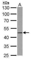
- Experimental details
- GFAP antibody detects GFAP protein by Western blot analysis.A. 50 µg mouse brain lysate/extract10 % SDS-PAGEGFAP antibody (GTX108711) dilution: 1:20000
- Validation comment
- WB
- Submitted by
- GeneTex (provider)
- Main image
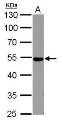
- Experimental details
- GFAP antibody detects GFAP protein by Western blot analysis.A. 50 µg rat brain lysate/extract10 % SDS-PAGEGFAP antibody (GTX108711) dilution: 1:20000
- Validation comment
- WB
- Submitted by
- GeneTex (provider)
- Main image
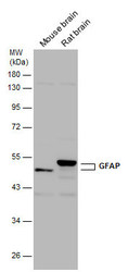
- Experimental details
- Various tissue extracts (50 ?g) were separated by 10% SDS-PAGE, and the membrane was blotted with GFAP antibody (GTX108711) diluted at 1:30000. The HRP-conjugated anti-rabbit IgG antibody (GTX213110-01) was used to detect the primary antibody.
- Submitted by
- GeneTex (provider)
- Main image
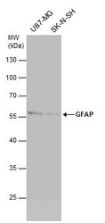
- Experimental details
- Various whole cell extracts (30 ?g) were separated by 10% SDS-PAGE, and the membrane was blotted with GFAP antibody (GTX108711) diluted at 1:20000. The HRP-conjugated anti-rabbit IgG antibody (GTX213110-01) was used to detect the primary antibody.
- Submitted by
- GeneTex (provider)
- Main image
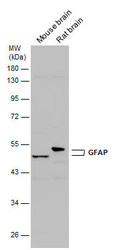
- Experimental details
- Various tissue extracts (50 ?g) were separated by 10% SDS-PAGE, and the membrane was blotted with GFAP antibody (GTX108711) diluted at 1:50000.
- Submitted by
- GeneTex (provider)
- Main image
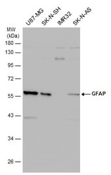
- Experimental details
- Various whole cell extracts (30 ?g) were separated by 10% SDS-PAGE, and the membrane was blotted with GFAP antibody (GTX108711) diluted at 1:20000. The HRP-conjugated anti-rabbit IgG antibody (GTX213110-01) was used to detect the primary antibody.
Supportive validation
- Submitted by
- GeneTex (provider)
- Main image

- Experimental details
- GFAP antibody detects GFAP protein at glia cells by immunofluorescent analysis.Sample: DIV9 rat E18 primary cortical neurons were fixed in 4% paraformaldehyde at RT for 15 min.Green: GFAP protein stained by GFAP antibody (GTX108711) diluted at 1:500.Red: beta Tubulin 3/ Tuj1, a neuron cell marker, stained by beta Tubulin 3/ Tuj1 antibody [GT11710] (GTX631836) diluted at 1:500.Blue: Fluoroshield with DAPI (GTX30920).
Supportive validation
- Submitted by
- GeneTex (provider)
- Main image
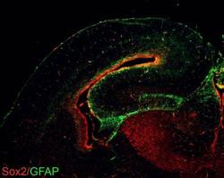
- Experimental details
- GFAP antibodies detects GFAP proteins on embryonic mouse brain by immunohistochemical analysis. Sample: Frozen section of embryonic mouse brain (mE18.5). Green: GFAP antibody (GTX108711) diluted at 1:1000. Red: Sox2 antibody [GT1352] (GTX627405) diluted at 1:500.
- Submitted by
- GeneTex (provider)
- Main image

- Experimental details
- GFAP antibody detects GFAP protein at cytoplasm in rat brain by immunohistochemical analysis. Sample: Paraffin-embedded rat brain. Green: GFAP antibody (GTX108711) diluted at 1:200. The signal was developed using goat anti-rabbit IgG antibody (Dylight488) (GTX213110-04).Blue: Nuclear staining with Hoechst 33342.
- Submitted by
- GeneTex (provider)
- Main image
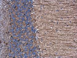
- Experimental details
- GFAP antibody detects GFAP protein at cytoplasm in rat brain by immunohistochemical analysis. Sample: Paraffin-embedded rat brain. GFAP antibody (GTX108711) diluted at 1:500.
- Submitted by
- GeneTex (provider)
- Main image
- Experimental details
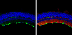
- GFAP antibody detects GFAP protein at retinal ganglion cell layer by immunohistochemical analysis.Sample: Frozen sectioned adult mouse retina. Green: GFAP protein stained by GFAP antibody (GTX108711) diluted at 1:250.Red: beta Tubulin 3/ TUJ1, stained by beta Tubulin 3/ TUJ1 antibody [GT11710] (GTX631836) diluted at 1:250.Blue: Fluoroshield with DAPI (GTX30920).
- Submitted by
- GeneTex (provider)
- Main image
- Experimental details
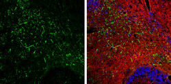
- GFAP antibody detects GFAP protein expression by immunohistochemical analysis.Sample: Frozen-sectioned adult mouse cerebellum. Green: GFAP protein stained by GFAP antibody (GTX108711) diluted at 1:250.Red: beta Tubulin 3/ TUJ1, stained by beta Tubulin 3/ TUJ1 antibody [GT11710] (GTX631836) diluted at 1:500.Blue: Fluoroshield with DAPI (GTX30920).
- Submitted by
- GeneTex (provider)
- Main image
- Experimental details
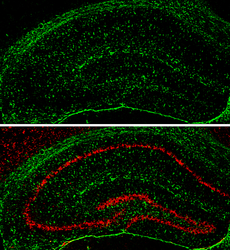
- GFAP antibody detects GFAP protein expression by immunohistochemical analysis.Sample: Frozen-sectioned adult mouse hippocampus. Green: GFAP protein stained by GFAP antibody (GTX108711) diluted at 1:250.Red: NeuN, stained by NeuN antibody [2Q158] (GTX30773) diluted at 1:500.
 Explore
 Validate
 Learn
Learn Western blot