Explore
 Validate
 Learn
Learn Western blot
Antibody data
- Antibody Data
- Antigen structure
- References [0]
- Comments [0]
- Validations
- Western blot [3]
- Immunohistochemistry [13]
Submit
Validation data
Reference
Comment
Report error
- Product number
- AMAb91033 - Provider product page

- Provider
- Atlas Antibodies
- Proper citation
- Atlas Antibodies Cat#AMAb91033, RRID:AB_2665775
- Product name
- Anti-GFAP
- Antibody type
- Monoclonal
- Reactivity
- Human, Mouse, Rat
- Host
- Mouse
- Conjugate
- Unconjugated
- Antigen sequence
LEGEENRITIPVQTFSNLQIRETSLDTKSVSEGHL
KRNIVVKTVEMRDGEVIKESKQEHKD- Epitope
- Binds to an epitope located within the peptide sequence PVQTFSNLQIRETSL as determined by overlapping synthetic peptides.
- Isotype
- IgG
- Antibody clone number
- CL2713
- Vial size
- 100 µl
- Storage
- Store at +4°C for short term storage. Long time storage is recommended at -20°C.
No comments: Submit comment
Enhanced validation
- Submitted by
- Atlas Antibodies (provider)
- Enhanced method
- Genetic validation
- Main image
- Experimental details
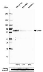
- Western blot analysis in U-87MG ATCC cells transfected with control siRNA, target specific siRNA probe #1 and #2, using Anti-GFAP antibody. Remaining relative intensity is presented. Loading control: Anti-GAPDH.
- Submitted by
- Atlas Antibodies (provider)
- Main image
- Experimental details
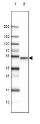
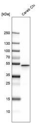
- Lane 1: Marker [kDa] 250, 130, 100, 70, 55, 35, 25, 15, 10Lane 2: Mouse Cerebral Cortex tissue

- Submitted by
- Atlas Antibodies (provider)
- Main image
- Experimental details
- Western blot analysis in mouse cerebral cortex tissue.

Enhanced validation
Supportive validation
- Submitted by
- Atlas Antibodies (provider)
- Enhanced method
- Orthogonal validation
- Main image
- Experimental details
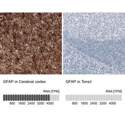
- Immunohistochemistry analysis in human cerebral cortex and tonsil tissues using AMAb91033 antibody. Corresponding GFAP RNA-seq data are presented for the same tissues.
- Sample type
- HUMAN

Supportive validation
- Submitted by
- Atlas Antibodies (provider)
- Main image
- Experimental details
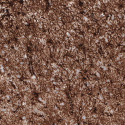
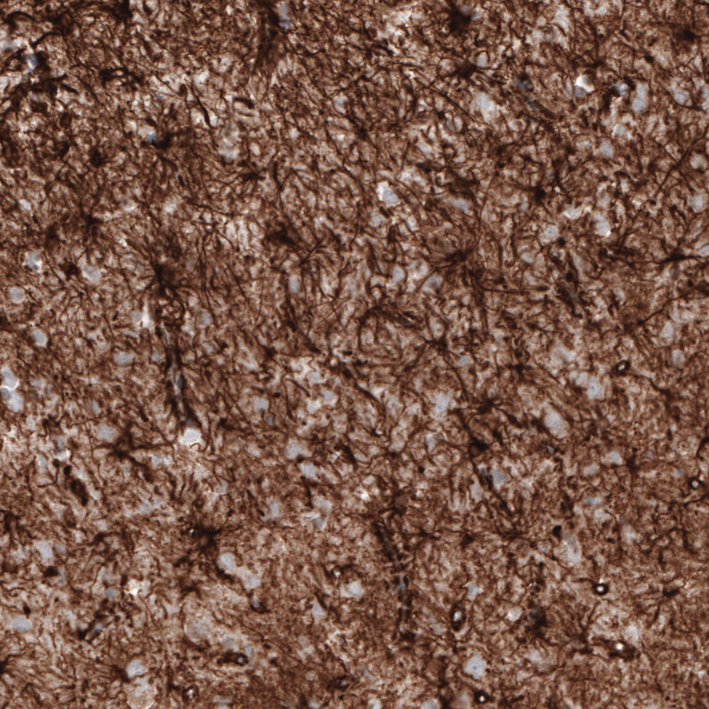
- Immunohistochemical staining of rat hippocampus shows strong immunoreactivity in astrocytes.
- Submitted by
- Atlas Antibodies (provider)
- Main image
- Experimental details
- Immunohistochemical staining of mouse cerebellum shows strong positivity in astrocytes.
- Submitted by
- Atlas Antibodies (provider)
- Main image
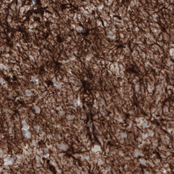
- Experimental details
- Immunohistochemical staining of human cerebral cortex shows strong immunoreactivity in astrocytes.
- Submitted by
- Atlas Antibodies (provider)
- Main image
- Experimental details
- Immunohistochemical staining of rat cerebellum shows strong positivity in astrocytes.

- Submitted by
- Atlas Antibodies (provider)
- Main image

- Experimental details
- Immunohistochemical staining of mouse hypothalamus shows strong immunoreactivity in astrocytes.

- Submitted by
- Atlas Antibodies (provider)
- Main image
- Experimental details
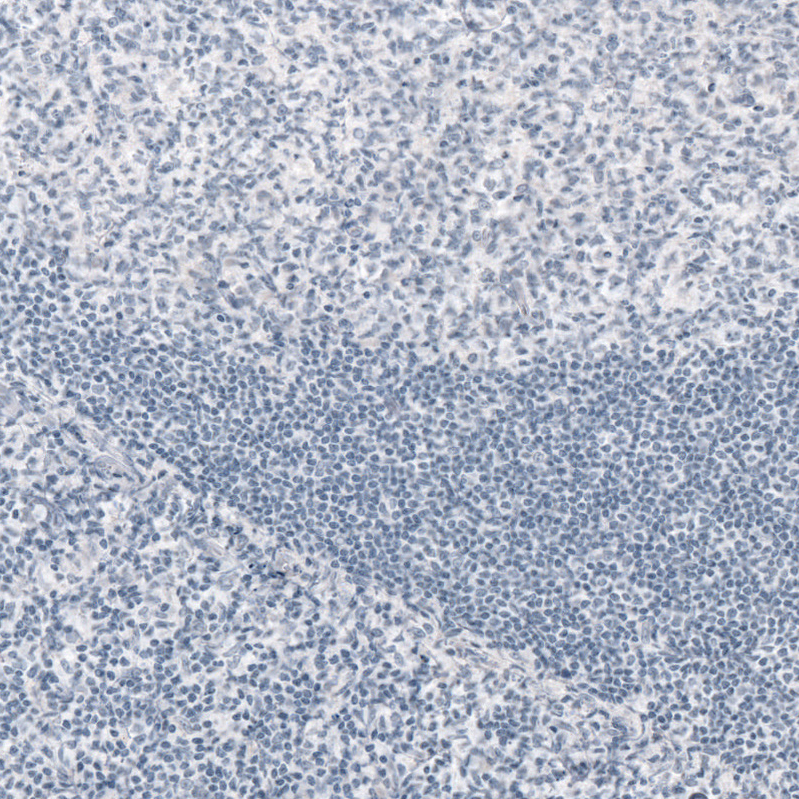
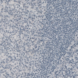
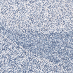
- Immunohistochemical staining of human tonsil shows absence of immunoreactivity (negative control).
- Submitted by
- Atlas Antibodies (provider)
- Main image
- Experimental details
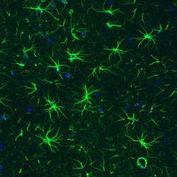
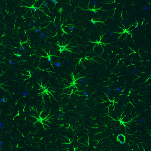
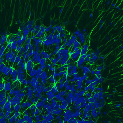
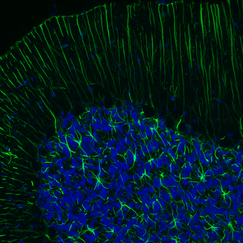
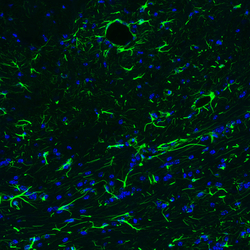
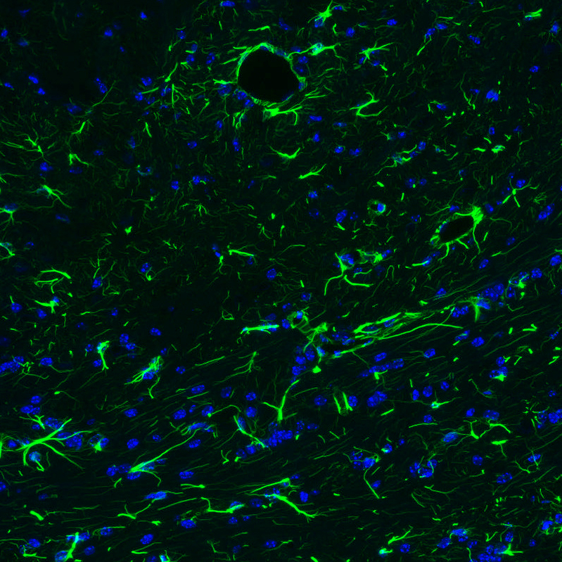
- Immunofluorescence staining of rat brain shows strong positivity in astrocytes in the hippocampus.

- Submitted by
- Atlas Antibodies (provider)
- Main image
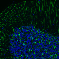
- Experimental details
- Immunofluorescence staining of rat brain shows strong positivity in astrocytes in the cerebellum.
- Submitted by
- Atlas Antibodies (provider)
- Main image
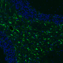
- Experimental details
- Immunofluorescence staining of mouse brain shows strong positivity in astrocytes in the cerebellum.
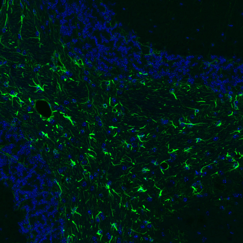
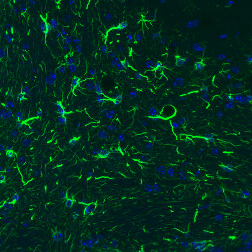
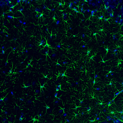
- Submitted by
- Atlas Antibodies (provider)
- Main image
- Experimental details
- Immunofluorescence staining of mouse brain shows strong positivity in astrocytes in the brainstem.
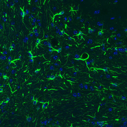
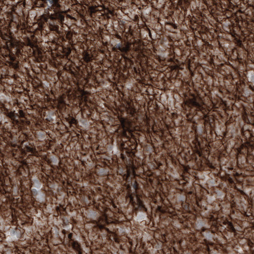
- Submitted by
- Atlas Antibodies (provider)
- Main image
- Experimental details
- Immunohistochemical staining of human cerebral cortex shows strong cytoplasmic positivity in astrocytes.
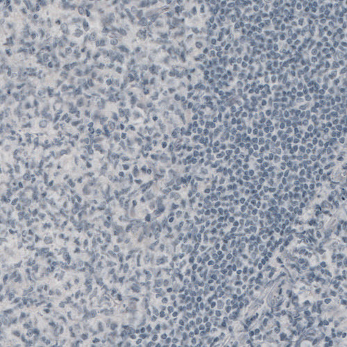
- Submitted by
- Atlas Antibodies (provider)
- Main image
- Experimental details
- Immunohistochemical staining of human tonsil shows no positivity in lymphoid cells as expected.