Antibody data
- Antibody Data
- Antigen structure
- References [54]
- Comments [0]
- Validations
- Western blot [2]
- Immunocytochemistry [4]
- Immunohistochemistry [5]
- Other assay [13]
Submit
Validation data
Reference
Comment
Report error
- Product number
- MA5-12023 - Provider product page

- Provider
- Invitrogen Antibodies
- Product name
- GFAP Monoclonal Antibody (ASTRO6)
- Antibody type
- Monoclonal
- Antigen
- Other
- Description
- MA5-12023 targets Glial Fibrillary Acidic Protein in IF, IHC (P), and WB applications and shows reactivity with Chicken, Human, Porcine, and Rat samples. The MA5-12023 immunogen is glial Fibrillary Acidic Protein. MA5-12023 was successfully used to detect astrocytes in E18 Sparague Dawley primary cortical cells.
- Reactivity
- Human, Rat, Chicken/Avian, Porcine
- Host
- Mouse
- Isotype
- IgG
- Antibody clone number
- ASTRO6
- Vial size
- 500 µL
- Concentration
- 200 mg/mL
- Storage
- 4° C
Submitted references Neurogenesis Is Reduced at 48 h in the Subventricular Zone Independent of Cell Death in a Piglet Model of Perinatal Hypoxia-Ischemia.
Epigenetic Studies in the Male APP/BIN1/COPS5 Triple-Transgenic Mouse Model of Alzheimer's Disease.
Detection and Functional Evaluation of the P2X7 Receptor in hiPSC Derived Neurons and Microglia-Like Cells.
Ultrarapid Inflammation of the Olfactory Bulb After Spinal Cord Injury: Protective Effects of the Granulocyte Colony-Stimulating Factor on Early Neurodegeneration in the Brain.
Phenotypic and functional comparison of rat enteric neural crest-derived cells during fetal and early-postnatal stages.
Decellularized spinal cord meninges extracellular matrix hydrogel that supports neurogenic differentiation and vascular structure formation.
SIX1/EYA1 are novel liver damage biomarkers in chronic hepatitis B and other liver diseases.
The epidermal growth factor receptor variant type III mutation frequently found in gliomas induces astrogenesis in human cerebral organoids.
Specific Frequency Electroacupuncture Stimulation Transiently Enhances the Permeability of the Blood-Brain Barrier and Induces Tight Junction Changes.
Herpes simplex virus type 1 infection leads to neurodevelopmental disorder-associated neuropathological changes.
Isolation and characterization of human optic nerve head astrocytes and lamina cribrosa cells.
Elevated protein synthesis in microglia causes autism-like synaptic and behavioral aberrations.
Adjuvant therapeutic potential of tonabersat in the standard treatment of glioblastoma: A preclinical F98 glioblastoma rat model study.
Morphine immunomodulation prolongs inflammatory and postoperative pain while the novel analgesic ZH853 accelerates recovery and protects against latent sensitization.
Mitochondrial Bioenergetics in Brain Following Ozone Exposure in Rats Maintained on Coconut, Fish and Olive Oil-Rich Diets.
Histopathological changes in the choroid plexus after traumatic brain injury in the rats: a histologic and immunohistochemical study.
A Single Primary Blast-Induced Traumatic Brain Injury in a Rodent Model Causes Cell-Type Dependent Increase in Nicotinamide Adenine Dinucleotide Phosphate Oxidase Isoforms in Vulnerable Brain Regions.
Interleukin-1β secreted from betanodavirus-infected microglia caused the death of neurons in giant grouper brains.
Neurotoxic reactive astrocytes are induced by activated microglia.
Reactive astrocyte COX2-PGE2 production inhibits oligodendrocyte maturation in neonatal white matter injury.
Lipocalin-2 as an Infection-Related Biomarker to Predict Clinical Outcome in Ischemic Stroke.
Recurrent intracranial neurenteric cyst with malignant transformation: A case report and literature review.
Curcumin ameliorates neuropathic pain by down-regulating spinal IL-1β via suppressing astroglial NALP1 inflammasome and JAK2-STAT3 signalling.
Systemic and cerebral iron homeostasis in ferritin knock-out mice.
Rapid generation of sub-type, region-specific neurons and neural networks from human pluripotent stem cell-derived neurospheres.
Spinal IL-33/ST2 Signaling Contributes to Neuropathic Pain via Neuronal CaMKII-CREB and Astroglial JAK2-STAT3 Cascades in Mice.
Electroacupuncture promotes proliferation of amplifying neural progenitors and preserves quiescent neural progenitors from apoptosis to alleviate depressive-like and anxiety-like behaviours.
Enhancement of Neural Stem Cells after Induction of Depression in Male Albino Rats (A histological & Immunohistochemical Study).
TDP-43 causes differential pathology in neuronal versus glial cells in the mouse brain.
Multipotent adult hippocampal progenitor cells maintained as neurospheres favor differentiation toward glial lineages.
Thymic stromal lymphopoietin is expressed in the intact central nervous system and upregulated in the myelin-degenerative central nervous system.
Fractalkine signaling and Tau hyper-phosphorylation are associated with autophagic alterations in lentiviral Tau and Aβ1-42 gene transfer models.
The multifaceted effects of agmatine on functional recovery after spinal cord injury through Modulations of BMP-2/4/7 expressions in neurons and glial cells.
Involvement of TREK-1 activity in astrocyte function and neuroprotection under simulated ischemia conditions.
Intrathecal lamotrigine attenuates mechanical allodynia and suppresses microglial and astrocytic activation in a rat model of spinal nerve ligation.
NDGA reduces secondary damage after spinal cord injury in rats via anti-inflammatory effects.
Development of a chemically extracted acellular muscle scaffold seeded with amniotic epithelial cells to promote spinal cord repair.
T cell-activation in neuromyelitis optica lesions plays a role in their formation.
Involvement of the spinal NALP1 inflammasome in neuropathic pain and aspirin-triggered-15-epi-lipoxin A4 induced analgesia.
Cdh1 inhibits reactive astrocyte proliferation after oxygen-glucose deprivation and reperfusion.
Changes in retinal aquaporin-9 (AQP9) expression in glaucoma.
Expression pattern of ataxia telangiectasia mutated (ATM), p53, Akt, and glycogen synthase kinase-3β in the striatum of rats treated with 3-nitropropionic acid.
Activation of Src family kinases in spinal microglia contributes to formalin-induced persistent pain state through p38 pathway.
Conditional Müllercell ablation causes independent neuronal and vascular pathologies in a novel transgenic model.
DLK1, delta-like 1 homolog (Drosophila), regulates tumor cell differentiation in vivo.
Multiple endocardial neurofibromas in a rosy-billed pochard (Netta peposaca).
NADPH oxidase expression in active multiple sclerosis lesions in relation to oxidative tissue damage and mitochondrial injury.
Genetic ablation of Pals1 in retinal progenitor cells models the retinal pathology of Leber congenital amaurosis.
Differentiated adipose-derived stem cells promote myelination and enhance functional recovery in a rat model of chronic denervation.
Changes in lipid-sensitive two-pore domain potassium channel TREK-1 expression and its involvement in astrogliosis following cerebral ischemia in rats.
The correlation between DTI parameters and levels of AQP-4 in the early phases of cerebral edema after hypoxic-ischemic/reperfusion injury in piglets.
Adsorption of mesenchymal stem cells and cortical neural stem cells on carbon nanotube/polycarbonate urethane.
CD8+ T-cell clones dominate brain infiltrates in Rasmussen encephalitis and persist in the periphery.
Embryonic porcine liver as a source for transplantation: advantage of intact liver implants over isolated hepatoblasts in overcoming homeostatic inhibition by the quiescent host liver.
Alonso-Alconada D, Gressens P, Golay X, Robertson NJ
Frontiers in pediatrics 2022;10:793189
Frontiers in pediatrics 2022;10:793189
Epigenetic Studies in the Male APP/BIN1/COPS5 Triple-Transgenic Mouse Model of Alzheimer's Disease.
Martínez-Iglesias O, Naidoo V, Carrera I, Cacabelos R
International journal of molecular sciences 2022 Feb 23;23(5)
International journal of molecular sciences 2022 Feb 23;23(5)
Detection and Functional Evaluation of the P2X7 Receptor in hiPSC Derived Neurons and Microglia-Like Cells.
Francistiová L, Vörös K, Lovász Z, Dinnyés A, Kobolák J
Frontiers in molecular neuroscience 2021;14:793769
Frontiers in molecular neuroscience 2021;14:793769
Ultrarapid Inflammation of the Olfactory Bulb After Spinal Cord Injury: Protective Effects of the Granulocyte Colony-Stimulating Factor on Early Neurodegeneration in the Brain.
Lin MS, Chiu IH, Lin CC
Frontiers in aging neuroscience 2021;13:701702
Frontiers in aging neuroscience 2021;13:701702
Phenotypic and functional comparison of rat enteric neural crest-derived cells during fetal and early-postnatal stages.
Tian DH, Qin CH, Xu WY, Pan WK, Zhao YY, Zheng BJ, Chen XL, Liu Y, Gao Y, Yu H
Neural regeneration research 2021 Nov;16(11):2310-2315
Neural regeneration research 2021 Nov;16(11):2310-2315
Decellularized spinal cord meninges extracellular matrix hydrogel that supports neurogenic differentiation and vascular structure formation.
Ozudogru E, Isik M, Eylem CC, Nemutlu E, Arslan YE, Derkus B
Journal of tissue engineering and regenerative medicine 2021 Nov;15(11):948-963
Journal of tissue engineering and regenerative medicine 2021 Nov;15(11):948-963
SIX1/EYA1 are novel liver damage biomarkers in chronic hepatitis B and other liver diseases.
Xu B, Yang Q, Tang Y, Tan Z, Fu H, Peng J, Xiang X, Gan L, Deng G, Mao Q, Xu PX, Jiang Y, Ding J
Annals of translational medicine 2021 Jun;9(12):992
Annals of translational medicine 2021 Jun;9(12):992
The epidermal growth factor receptor variant type III mutation frequently found in gliomas induces astrogenesis in human cerebral organoids.
Kim HM, Lee SH, Lim J, Yoo J, Hwang DY
Cell proliferation 2021 Feb;54(2):e12965
Cell proliferation 2021 Feb;54(2):e12965
Specific Frequency Electroacupuncture Stimulation Transiently Enhances the Permeability of the Blood-Brain Barrier and Induces Tight Junction Changes.
Zhang S, Gong P, Zhang J, Mao X, Zhao Y, Wang H, Gan L, Lin X
Frontiers in neuroscience 2020;14:582324
Frontiers in neuroscience 2020;14:582324
Herpes simplex virus type 1 infection leads to neurodevelopmental disorder-associated neuropathological changes.
Qiao H, Guo M, Shang J, Zhao W, Wang Z, Liu N, Li B, Zhou Y, Wu Y, Chen P
PLoS pathogens 2020 Oct;16(10):e1008899
PLoS pathogens 2020 Oct;16(10):e1008899
Isolation and characterization of human optic nerve head astrocytes and lamina cribrosa cells.
Lopez NN, Clark AF, Tovar-Vidales T
Experimental eye research 2020 Aug;197:108103
Experimental eye research 2020 Aug;197:108103
Elevated protein synthesis in microglia causes autism-like synaptic and behavioral aberrations.
Xu ZX, Kim GH, Tan JW, Riso AE, Sun Y, Xu EY, Liao GY, Xu H, Lee SH, Do NY, Lee CH, Clipperton-Allen AE, Kwon S, Page DT, Lee KJ, Xu B
Nature communications 2020 Apr 14;11(1):1797
Nature communications 2020 Apr 14;11(1):1797
Adjuvant therapeutic potential of tonabersat in the standard treatment of glioblastoma: A preclinical F98 glioblastoma rat model study.
De Meulenaere V, Bonte E, Verhoeven J, Kalala Okito JP, Pieters L, Vral A, De Wever O, Leybaert L, Goethals I, Vanhove C, Descamps B, Deblaere K
PloS one 2019;14(10):e0224130
PloS one 2019;14(10):e0224130
Morphine immunomodulation prolongs inflammatory and postoperative pain while the novel analgesic ZH853 accelerates recovery and protects against latent sensitization.
Feehan AK, Zadina JE
Journal of neuroinflammation 2019 May 21;16(1):100
Journal of neuroinflammation 2019 May 21;16(1):100
Mitochondrial Bioenergetics in Brain Following Ozone Exposure in Rats Maintained on Coconut, Fish and Olive Oil-Rich Diets.
Valdez MC, Freeborn D, Valdez JM, Johnstone AFM, Snow SJ, Tennant AH, Kodavanti UP, Kodavanti PRS
International journal of molecular sciences 2019 Dec 13;20(24)
International journal of molecular sciences 2019 Dec 13;20(24)
Histopathological changes in the choroid plexus after traumatic brain injury in the rats: a histologic and immunohistochemical study.
Özevren H, Deveci E, Tuncer MC
Folia morphologica 2018;77(4):642-648
Folia morphologica 2018;77(4):642-648
A Single Primary Blast-Induced Traumatic Brain Injury in a Rodent Model Causes Cell-Type Dependent Increase in Nicotinamide Adenine Dinucleotide Phosphate Oxidase Isoforms in Vulnerable Brain Regions.
Rama Rao KV, Iring S, Younger D, Kuriakose M, Skotak M, Alay E, Gupta RK, Chandra N
Journal of neurotrauma 2018 Sep 1;35(17):2077-2090
Journal of neurotrauma 2018 Sep 1;35(17):2077-2090
Interleukin-1β secreted from betanodavirus-infected microglia caused the death of neurons in giant grouper brains.
Chiang YH, Wu YC, Chi SC
Developmental and comparative immunology 2017 May;70:19-26
Developmental and comparative immunology 2017 May;70:19-26
Neurotoxic reactive astrocytes are induced by activated microglia.
Liddelow SA, Guttenplan KA, Clarke LE, Bennett FC, Bohlen CJ, Schirmer L, Bennett ML, Münch AE, Chung WS, Peterson TC, Wilton DK, Frouin A, Napier BA, Panicker N, Kumar M, Buckwalter MS, Rowitch DH, Dawson VL, Dawson TM, Stevens B, Barres BA
Nature 2017 Jan 26;541(7638):481-487
Nature 2017 Jan 26;541(7638):481-487
Reactive astrocyte COX2-PGE2 production inhibits oligodendrocyte maturation in neonatal white matter injury.
Shiow LR, Favrais G, Schirmer L, Schang AL, Cipriani S, Andres C, Wright JN, Nobuta H, Fleiss B, Gressens P, Rowitch DH
Glia 2017 Dec;65(12):2024-2037
Glia 2017 Dec;65(12):2024-2037
Lipocalin-2 as an Infection-Related Biomarker to Predict Clinical Outcome in Ischemic Stroke.
Hochmeister S, Engel O, Adzemovic MZ, Pekar T, Kendlbacher P, Zeitelhofer M, Haindl M, Meisel A, Fazekas F, Seifert-Held T
PloS one 2016;11(5):e0154797
PloS one 2016;11(5):e0154797
Recurrent intracranial neurenteric cyst with malignant transformation: A case report and literature review.
Yang Y, Fang J, Li DA, Wang L, Ji N, Zhang J
Oncology letters 2016 May;11(5):3395-3402
Oncology letters 2016 May;11(5):3395-3402
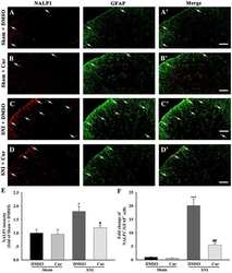
Curcumin ameliorates neuropathic pain by down-regulating spinal IL-1β via suppressing astroglial NALP1 inflammasome and JAK2-STAT3 signalling.
Liu S, Li Q, Zhang MT, Mao-Ying QL, Hu LY, Wu GC, Mi WL, Wang YQ
Scientific reports 2016 Jul 6;6:28956
Scientific reports 2016 Jul 6;6:28956
Systemic and cerebral iron homeostasis in ferritin knock-out mice.
Li W, Garringer HJ, Goodwin CB, Richine B, Acton A, VanDuyn N, Muhoberac BB, Irimia-Dominguez J, Chan RJ, Peacock M, Nass R, Ghetti B, Vidal R
PloS one 2015;10(1):e0117435
PloS one 2015;10(1):e0117435
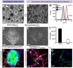
Rapid generation of sub-type, region-specific neurons and neural networks from human pluripotent stem cell-derived neurospheres.
Begum AN, Guoynes C, Cho J, Hao J, Lutfy K, Hong Y
Stem cell research 2015 Nov;15(3):731-741
Stem cell research 2015 Nov;15(3):731-741
Spinal IL-33/ST2 Signaling Contributes to Neuropathic Pain via Neuronal CaMKII-CREB and Astroglial JAK2-STAT3 Cascades in Mice.
Liu S, Mi WL, Li Q, Zhang MT, Han P, Hu S, Mao-Ying QL, Wang YQ
Anesthesiology 2015 Nov;123(5):1154-69
Anesthesiology 2015 Nov;123(5):1154-69
Electroacupuncture promotes proliferation of amplifying neural progenitors and preserves quiescent neural progenitors from apoptosis to alleviate depressive-like and anxiety-like behaviours.
Yang L, Yue N, Zhu X, Han Q, Li B, Liu Q, Wu G, Yu J
Evidence-based complementary and alternative medicine : eCAM 2014;2014:872568
Evidence-based complementary and alternative medicine : eCAM 2014;2014:872568
Enhancement of Neural Stem Cells after Induction of Depression in Male Albino Rats (A histological & Immunohistochemical Study).
Kamel Ismail ZM, Morcos MA, Eldin Mohammad MD, Gamal Aboulkhair A
International journal of stem cells 2014 Nov;7(2):70-8
International journal of stem cells 2014 Nov;7(2):70-8
TDP-43 causes differential pathology in neuronal versus glial cells in the mouse brain.
Yan S, Wang CE, Wei W, Gaertig MA, Lai L, Li S, Li XJ
Human molecular genetics 2014 May 15;23(10):2678-93
Human molecular genetics 2014 May 15;23(10):2678-93
Multipotent adult hippocampal progenitor cells maintained as neurospheres favor differentiation toward glial lineages.
Oh J, Daniels GJ, Chiou LS, Ye EA, Jeong YS, Sakaguchi DS
Biotechnology journal 2014 Jul;9(7):921-33
Biotechnology journal 2014 Jul;9(7):921-33
Thymic stromal lymphopoietin is expressed in the intact central nervous system and upregulated in the myelin-degenerative central nervous system.
Kitic M, Wimmer I, Adzemovic M, Kögl N, Rudel A, Lassmann H, Bradl M
Glia 2014 Jul;62(7):1066-74
Glia 2014 Jul;62(7):1066-74
Fractalkine signaling and Tau hyper-phosphorylation are associated with autophagic alterations in lentiviral Tau and Aβ1-42 gene transfer models.
Hebron ML, Algarzae NK, Lonskaya I, Moussa C
Experimental neurology 2014 Jan;251:127-38
Experimental neurology 2014 Jan;251:127-38
The multifaceted effects of agmatine on functional recovery after spinal cord injury through Modulations of BMP-2/4/7 expressions in neurons and glial cells.
Park YM, Lee WT, Bokara KK, Seo SK, Park SH, Kim JH, Yenari MA, Park KA, Lee JE
PloS one 2013;8(1):e53911
PloS one 2013;8(1):e53911
Involvement of TREK-1 activity in astrocyte function and neuroprotection under simulated ischemia conditions.
Wu X, Liu Y, Chen X, Sun Q, Tang R, Wang W, Yu Z, Xie M
Journal of molecular neuroscience : MN 2013 Mar;49(3):499-506
Journal of molecular neuroscience : MN 2013 Mar;49(3):499-506
Intrathecal lamotrigine attenuates mechanical allodynia and suppresses microglial and astrocytic activation in a rat model of spinal nerve ligation.
Choi YS, Jun IG, Kim SH, Park JY
Yonsei medical journal 2013 Mar 1;54(2):321-9
Yonsei medical journal 2013 Mar 1;54(2):321-9
NDGA reduces secondary damage after spinal cord injury in rats via anti-inflammatory effects.
Xue H, Zhang XY, Liu JM, Song Y, Liu TT, Chen D
Brain research 2013 Jun 21;1516:83-92
Brain research 2013 Jun 21;1516:83-92
Development of a chemically extracted acellular muscle scaffold seeded with amniotic epithelial cells to promote spinal cord repair.
Xue H, Zhang XY, Liu JM, Song Y, Li YF, Chen D
Journal of biomedical materials research. Part A 2013 Jan;101(1):145-56
Journal of biomedical materials research. Part A 2013 Jan;101(1):145-56
T cell-activation in neuromyelitis optica lesions plays a role in their formation.
Pohl M, Kawakami N, Kitic M, Bauer J, Martins R, Fischer MT, Machado-Santos J, Mader S, Ellwart JW, Misu T, Fujihara K, Wekerle H, Reindl M, Lassmann H, Bradl M
Acta neuropathologica communications 2013 Dec 24;1:85
Acta neuropathologica communications 2013 Dec 24;1:85
Involvement of the spinal NALP1 inflammasome in neuropathic pain and aspirin-triggered-15-epi-lipoxin A4 induced analgesia.
Li Q, Tian Y, Wang ZF, Liu SB, Mi WL, Ma HJ, Wu GC, Wang J, Yu J, Wang YQ
Neuroscience 2013 Dec 19;254:230-40
Neuroscience 2013 Dec 19;254:230-40
Cdh1 inhibits reactive astrocyte proliferation after oxygen-glucose deprivation and reperfusion.
Qiu J, Zhang C, Lv Y, Zhang Y, Zhu C, Wang X, Yao W
Neurochemistry international 2013 Aug;63(2):87-92
Neurochemistry international 2013 Aug;63(2):87-92
Changes in retinal aquaporin-9 (AQP9) expression in glaucoma.
Yang MH, Dibas A, Tyan YC
Bioscience reports 2013 Apr 23;33(2)
Bioscience reports 2013 Apr 23;33(2)
Expression pattern of ataxia telangiectasia mutated (ATM), p53, Akt, and glycogen synthase kinase-3β in the striatum of rats treated with 3-nitropropionic acid.
Duran-Vilaregut J, Manich G, Del Valle J, Camins A, Pallàs M, Vilaplana J, Pelegrí C
Journal of neuroscience research 2012 Sep;90(9):1803-13
Journal of neuroscience research 2012 Sep;90(9):1803-13
Activation of Src family kinases in spinal microglia contributes to formalin-induced persistent pain state through p38 pathway.
Tan YH, Li K, Chen XY, Cao Y, Light AR, Fu KY
The journal of pain 2012 Oct;13(10):1008-15
The journal of pain 2012 Oct;13(10):1008-15
Conditional Müllercell ablation causes independent neuronal and vascular pathologies in a novel transgenic model.
Shen W, Fruttiger M, Zhu L, Chung SH, Barnett NL, Kirk JK, Lee S, Coorey NJ, Killingsworth M, Sherman LS, Gillies MC
The Journal of neuroscience : the official journal of the Society for Neuroscience 2012 Nov 7;32(45):15715-27
The Journal of neuroscience : the official journal of the Society for Neuroscience 2012 Nov 7;32(45):15715-27
DLK1, delta-like 1 homolog (Drosophila), regulates tumor cell differentiation in vivo.
Begum A, Kim Y, Lin Q, Yun Z
Cancer letters 2012 May 1;318(1):26-33
Cancer letters 2012 May 1;318(1):26-33
Multiple endocardial neurofibromas in a rosy-billed pochard (Netta peposaca).
Miller AD, Baitchman EJ, Masek-Hammerman K
Journal of veterinary diagnostic investigation : official publication of the American Association of Veterinary Laboratory Diagnosticians, Inc 2012 Mar;24(2):408-11
Journal of veterinary diagnostic investigation : official publication of the American Association of Veterinary Laboratory Diagnosticians, Inc 2012 Mar;24(2):408-11
NADPH oxidase expression in active multiple sclerosis lesions in relation to oxidative tissue damage and mitochondrial injury.
Fischer MT, Sharma R, Lim JL, Haider L, Frischer JM, Drexhage J, Mahad D, Bradl M, van Horssen J, Lassmann H
Brain : a journal of neurology 2012 Mar;135(Pt 3):886-99
Brain : a journal of neurology 2012 Mar;135(Pt 3):886-99
Genetic ablation of Pals1 in retinal progenitor cells models the retinal pathology of Leber congenital amaurosis.
Cho SH, Kim JY, Simons DL, Song JY, Le JH, Swindell EC, Jamrich M, Wu SM, Kim S
Human molecular genetics 2012 Jun 15;21(12):2663-76
Human molecular genetics 2012 Jun 15;21(12):2663-76
Differentiated adipose-derived stem cells promote myelination and enhance functional recovery in a rat model of chronic denervation.
Tomita K, Madura T, Mantovani C, Terenghi G
Journal of neuroscience research 2012 Jul;90(7):1392-402
Journal of neuroscience research 2012 Jul;90(7):1392-402
Changes in lipid-sensitive two-pore domain potassium channel TREK-1 expression and its involvement in astrogliosis following cerebral ischemia in rats.
Wang M, Song J, Xiao W, Yang L, Yuan J, Wang W, Yu Z, Xie M
Journal of molecular neuroscience : MN 2012 Feb;46(2):384-92
Journal of molecular neuroscience : MN 2012 Feb;46(2):384-92
The correlation between DTI parameters and levels of AQP-4 in the early phases of cerebral edema after hypoxic-ischemic/reperfusion injury in piglets.
Wang H, Wang X, Guo Q
Pediatric radiology 2012 Aug;42(8):992-9
Pediatric radiology 2012 Aug;42(8):992-9
Adsorption of mesenchymal stem cells and cortical neural stem cells on carbon nanotube/polycarbonate urethane.
Nho Y, Kim JY, Khang D, Webster TJ, Lee JE
Nanomedicine (London, England) 2010 Apr;5(3):409-17
Nanomedicine (London, England) 2010 Apr;5(3):409-17
CD8+ T-cell clones dominate brain infiltrates in Rasmussen encephalitis and persist in the periphery.
Schwab N, Bien CG, Waschbisch A, Becker A, Vince GH, Dornmair K, Wiendl H
Brain : a journal of neurology 2009 May;132(Pt 5):1236-46
Brain : a journal of neurology 2009 May;132(Pt 5):1236-46
Embryonic porcine liver as a source for transplantation: advantage of intact liver implants over isolated hepatoblasts in overcoming homeostatic inhibition by the quiescent host liver.
Katchman H, Tal O, Eventov-Friedman S, Shezen E, Aronovich A, Tchorsh D, Cohen S, Shtabsky A, Hecht G, Dekel B, Freud E, Reisner Y
Stem cells (Dayton, Ohio) 2008 May;26(5):1347-55
Stem cells (Dayton, Ohio) 2008 May;26(5):1347-55
No comments: Submit comment
Supportive validation
- Submitted by
- Invitrogen Antibodies (provider)
- Main image

- Experimental details
- Western blot of Glial Fibrillary Acidic Protein using Glial Fibrillary Acidic Protein Monoclonal Antibody (Product # MA5-12023) on IMR-5 Cells.
- Submitted by
- Invitrogen Antibodies (provider)
- Main image

- Experimental details
- Western blot was performed using Anti-GFAP Monoclonal Antibody (ASTRO6), (Product # MA5-12023) and a 50 kDa band corresponding to GFAP was observed in the tissues tested along with Mouse IgG at ~25 kDa. Whole cell extracts (30 µg lysate) of Mouse brain (Lane 1), Rat brain (Lane 2), Mouse liver (Lane 3), and Rat liver (Lane 4) were electrophoresed using Novex® NuPAGE® 4-12 % Bis-Tris gel (Product # NP0321BOX). Resolved proteins were then transferred onto a nitrocellulose membrane (Product # IB23001) by iBlot® 2 Dry Blotting System (Product # IB21001). The blot was probed with the primary antibody (0.2 ug/ml) and detected by chemiluminescence with Goat anti-Mouse IgG (H+L), Superclonal™ Recombinant Secondary Antibody, HRP conjugate (Product # A28177, 1:4000 dilution) using the iBright FL 1000 (Product # A32752). Chemiluminescent detection was performed using Novex® ECL Chemiluminescent Substrate Reagent Kit (Product # WP20005).
Supportive validation
- Submitted by
- Invitrogen Antibodies (provider)
- Main image

- Experimental details
- Immunofluorescent analysis of GFAP (green) showing staining in the in the cytoplasm of SK-N-MC cells (right) compared to a negative control without primary antibody (left). Formalin-fixed cells were permeabilized with 0.1% Triton X-100 in TBS for 5-10 minutes and blocked with 3% BSA-PBS for 30 minutes at room temperature. Cells were probed with a GFAP monoclonal antibody (Product # MA5-12023) in 3% BSA-PBS at a dilution of 1:100 and incubated overnight at 4ºC in a humidified chamber. Cells were washed with PBST and incubated with a DyLight-conjugated secondary antibody in PBS at room temperature in the dark. F-actin (red) was stained with a fluorescent red phalloidin and nuclei (blue) were stained with Hoechst or DAPI. Images were taken at a magnification of 60x.
- Submitted by
- Invitrogen Antibodies (provider)
- Main image

- Experimental details
- Immunofluorescent analysis of glial fibrillary acidic protein (GFAP) in E18 Sparague Dawley primary cortical neuronal cells containing astrocytes. The cells were fixed with 4% formaldehyde for 15 mins, permeabilized with 0.25% Triton X-100 in PBS for 10 mins, and blocked with 3% BSA in PBS for 30 mins at RT. Cells were stained with a GFAP mouse monoclonal antibody (Product # MA5-12023) at a dilution of 1:200 in 3% BSA in PBS for 1 hr at RT, and then incubated with Invitrogen AlexaFluor 488 Plus goat anti-mouse IgG secondary antibody (Product # A32723) at a dilution of 1:1000 for 1 hr at RT. Nuclei were stained with Hoechst 33342 (Product # H3570). The image contains overlay of GFAP (green) and nuclei (blue). Images were taken on a Zeiss LSM 710 confocal microscope at 40X magnification.
- Submitted by
- Invitrogen Antibodies (provider)
- Main image

- Experimental details
- Immunofluorescent analysis of GFAP (green) showing staining in the in the cytoplasm of SK-N-MC cells (right) compared to a negative control without primary antibody (left). Formalin-fixed cells were permeabilized with 0.1% Triton X-100 in TBS for 5-10 minutes and blocked with 3% BSA-PBS for 30 minutes at room temperature. Cells were probed with a GFAP monoclonal antibody (Product # MA5-12023) in 3% BSA-PBS at a dilution of 1:100 and incubated overnight at 4ºC in a humidified chamber. Cells were washed with PBST and incubated with a DyLight-conjugated secondary antibody in PBS at room temperature in the dark. F-actin (red) was stained with a fluorescent red phalloidin and nuclei (blue) were stained with Hoechst or DAPI. Images were taken at a magnification of 60x.
- Submitted by
- Invitrogen Antibodies (provider)
- Main image

- Experimental details
- Immunofluorescent analysis of GFAP (green) showing staining in the in the cytoplasm of U251 cells (right) compared to a negative control without primary antibody (left). Formalin-fixed cells were permeabilized with 0.1% Triton X-100 in TBS for 5-10 minutes and blocked with 3% BSA-PBS for 30 minutes at room temperature. Cells were probed with a GFAP monoclonal antibody (Product # MA5-12023) in 3% BSA-PBS at a dilution of 1:100 and incubated overnight at 4ºC in a humidified chamber. Cells were washed with PBST and incubated with a DyLight-conjugated secondary antibody in PBS at room temperature in the dark. F-actin (red) was stained with a fluorescent red phalloidin and nuclei (blue) were stained with Hoechst or DAPI. Images were taken at a magnification of 60x.
Supportive validation
- Submitted by
- Invitrogen Antibodies (provider)
- Main image

- Experimental details
- Formalin-fixed, paraffin-embedded human brain stained with GFAP antibody using peroxidase-conjugate and DAB chromogen. Note cytoplasmic staining of astrocytes.
- Submitted by
- Invitrogen Antibodies (provider)
- Main image

- Experimental details
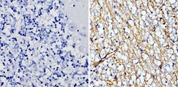
- Immunohistochemistry analysis of GFAP showing staining in the cytoplasm of paraffin-embedded human glioma (right) compared to a negative control without primary antibody (left). To expose target proteins, antigen retrieval was performed using 10mM sodium citrate (pH 6.0), microwaved for 8-15 min. Following antigen retrieval, tissues were blocked in 3% H2O2-methanol for 15 min at room temperature, washed with ddH2O and PBS, and then probed with a GFAP monoclonal antibody (Product # MA5-12023) diluted in 3% BSA-PBS at a dilution of 1:100 overnight at 4°C in a humidified chamber. Tissues were washed extensively in PBST and detection was performed using an HRP-conjugated secondary antibody followed by colorimetric detection using a DAB kit. Tissues were counterstained with hematoxylin and dehydrated with ethanol and xylene to prep for mounting.
- Submitted by
- Invitrogen Antibodies (provider)
- Main image

- Experimental details
- Immunohistochemistry analysis of GFAP showing staining in the cytoplasm of paraffin-embedded human cerebellum tissue (right) compared to a negative control without primary antibody (left). To expose target proteins, antigen retrieval was performed using 10mM sodium citrate (pH 6.0), microwaved for 8-15 min. Following antigen retrieval, tissues were blocked in 3% H2O2-methanol for 15 min at room temperature, washed with ddH2O and PBS, and then probed with a GFAP monoclonal antibody (Product # MA5-12023) diluted in 3% BSA-PBS at a dilution of 1:200 overnight at 4°C in a humidified chamber. Tissues were washed extensively in PBST and detection was performed using an HRP-conjugated secondary antibody followed by colorimetric detection using a DAB kit. Tissues were counterstained with hematoxylin and dehydrated with ethanol and xylene to prep for mounting.
- Submitted by
- Invitrogen Antibodies (provider)
- Main image

- Experimental details
- Immunohistochemistry analysis of GFAP showing staining in the cytoplasm of paraffin-embedded rat brain tissue (right) compared to a negative control without primary antibody (left). To expose target proteins, antigen retrieval was performed using 10mM sodium citrate (pH 6.0), microwaved for 8-15 min. Following antigen retrieval, tissues were blocked in 3% H2O2-methanol for 15 min at room temperature, washed with ddH2O and PBS, and then probed with a GFAP monoclonal antibody (Product # MA5-12023) diluted in 3% BSA-PBS at a dilution of 1:200 overnight at 4°C in a humidified chamber. Tissues were washed extensively in PBST and detection was performed using an HRP-conjugated secondary antibody followed by colorimetric detection using a DAB kit. Tissues were counterstained with hematoxylin and dehydrated with ethanol and xylene to prep for mounting.
- Submitted by
- Invitrogen Antibodies (provider)
- Main image

- Experimental details
- Immunofluorescent analysis of GFAP in human iPSC-derived astrocytes. The cells were fixed with 4% paraformaldehyde for 15 min at room temperature, and then permeabilized and blocked with 0.25% Triton X-100 and 10% donkey serum in PBS for 20 min. Samples were then incubated with a Mouse GFAP monoclonal antibody (green; Product # MA5-12023) at a dilution of 1:500 in PBS containing 0.25% Triton X-100 and 10% donkey serum at 48304C overnight, followed by incubation with Donkey anti-Mouse Alexa Fluor 488 (Product # R37114) at a dilution of 1:1,000 as well as DAPI (blue; 1:25,000) in PBS containing 0.25% Triton X-100 and 10% donkey serum at room temperature for 1 hour. Images were taken at 40X magnification. Data courtesy of Dr. Zhexing Wen at Emory University.
Supportive validation
- Submitted by
- Invitrogen Antibodies (provider)
- Main image

- Experimental details
- NULL
- Submitted by
- Invitrogen Antibodies (provider)
- Main image

- Experimental details
- NULL
- Submitted by
- Invitrogen Antibodies (provider)
- Main image

- Experimental details
- NULL
- Submitted by
- Invitrogen Antibodies (provider)
- Main image

- Experimental details
- NULL
- Submitted by
- Invitrogen Antibodies (provider)
- Main image

- Experimental details
- NULL
- Submitted by
- Invitrogen Antibodies (provider)
- Main image

- Experimental details
- Fig 8 HSV-1 infection accelerated the astrocytes activation in neuroepithelial buds and cerebral organoids. (A) Immunofluorescence staining for the astrocyte marker (GFAP) in neuroepithelial buds at the day 18 after 3 days with HSV-1 infection. The images were taken Images were captured on a Leica TCS SP8 STED confocal microscope. (B) Relative fluorescence intensity statistics of GFAP expression were shown in different groups. (C) Detection of GFAP mRNA expression in neuroepithelial buds (n = 4). Data represent the mean +- SEM. **p < 0.01 by Student's t test. (D) The sample images of immunostaining for GFAP in the cerebral organoids at the day 45 for 3 days with HSV-1 infection (Mock, Low infection, and High infection). Scale bars: 25 mum. (E) The relative fluorescence intensity statistics of GFAP expressions were shown in different groups. Data represent the mean +- SEM from three experiments. **p < 0.01 by ANOVA.
- Submitted by
- Invitrogen Antibodies (provider)
- Main image

- Experimental details
- FIGURE 2 Spinal cord injury (SCI) can activate the astrocytes in the olfactory bulb at 8 h post-spinal cord hemisection injury in mice. Representative images of GFAP-stained sections of the olfactory bulbs of the sham control (B) , SCI (C) , SCI + granulocyte colony-stimulating factor (G-CSF) i.p., and SCI + G-CSF oral groups. (A) Schematic illustration of the olfactory bulb (marked with dashed box) of all groups subjected to mRNA, protein, and immunofluorescence analyses. The SCI group exhibited a higher number of GFAP-positive cells [as indicated by arrow in panel (C) ] than the sham control group (B) . This indicated astrocytic activation and potential astrocyte-mediated inflammatory responses in the olfactory bulb at 8 h post-SCI. The immunofluorescence intensity of GFAP significantly decreased in the SCI + G-CSF i.p. [as indicated by arrow in panel (D) ] and SCI + G-CSF oral groups [as indicated by arrow in panel (E) ] [ (B-C) magnification 400x]. (E) Vertical bars indicate the mean +- standard error of mean) number of GFAP-stained cells in each group ( n = 3). *** P < 0.001 and ### P < 0.001.
- Submitted by
- Invitrogen Antibodies (provider)
- Main image

- Experimental details
- FIGURE 3 Neuroinflammation in the mouse olfactory bulb at 8 h post-spinal cord injury (SCI). (A-C) The mRNA expression levels of IL-1beta (A) , IL-6 (B) , and GFAP (C) in the olfactory bulb of the sham control, SCI, SCI + granulocyte colony-stimulating factor (G-CSF) i.p., and SCI + G-CSF oral groups at 8 h post-SCI. Vertical bars indicate the mean +- standard error of the mean (SEM) ( n = 6 for each group). * P < 0.05, *** P < 0.001, ## P < 0.01, and ### P < 0.001. (D-F) The protein expression levels of IL-1beta (D) , IL-6 (E) , and GFAP (F) in the olfactory bulb of the four experimental groups at 8 h post-SCI. Representative immunoblots of IL-1beta, IL-6, GFAP, and beta-actin (internal control) are shown in the upper panel. The lower panel indicates the ratio of target protein band intensity to beta-actin protein band intensity relative to the control group (mean +- SEM). G-CSF mitigates SCI-induced neuroinflammation in the olfactory bulb as evidenced by the decreased expression of IL-1beta, IL-6, and GFAP. Vertical bars indicate mean +- SEM ( n = 6 for each group). * P < 0.05, ** P < 0.01, and ## P < 0.01.
- Submitted by
- Invitrogen Antibodies (provider)
- Main image

- Experimental details
- 5 FIGURE Characterization of the neuro-inductive potential of meninges-derived hydrogel (MeninGEL). (a) Macroscopic images of MeninGELs after 10-day culture. (b) Inverted images of human mesenchymal stem cells (hMSCs) cultured in MeninGEL (10 mg mL -1 ) and Matrigel TM (10 mg mL -1 ) within growth medium and N2B27. (c) DAPI staining of emerged micro-tissues. (d) Immuno/histochemistry for hMSCs cultured within MeninGEL (10 mg mL -1 ) and Matrigel TM : hematoxylin and eosin, Nestin, MAP2, and GFAP staining. Gene expression study for Nestin (e), MAP2 (f), and GFAP (g) ( n = 3, * p < 0.05, ** p > 0.05)
- Submitted by
- Invitrogen Antibodies (provider)
- Main image

- Experimental details
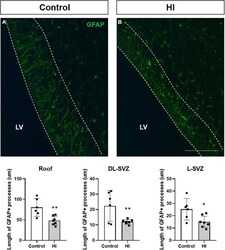
- FIGURE 5 Fluorescent microphotographs of GFAP immune-stained samples from control and HI piglets. GFAP reveals the length of the processes of radial-glia/neural stem cells in the SVZ (A) . HI [ (B) and graph] significantly reduced the length of GFAP + processes in the three subareas of the SVZ: roof ( ** p < 0.01), DL-SVZ ( ** p < 0.01), and L-SVZ (* p < 0.05). LV, lateral ventricle; SVZ, subventricular zone; CDT, caudate nucleus; PvWM, periventricular white matter. Original magnification 400x. Scale bar: 100 mum.
- Submitted by
- Invitrogen Antibodies (provider)
- Main image

- Experimental details
- Fig. 6. Role of the neuronal calcium-calmodulin-dependent kinase II (CaMKII)-cyclic adenosine monophosphate response element-binding protein (CREB) cascade in spinal interleukin (IL)-33&solST2 signaling-mediated neuropathic pain. ( A and B ) Western blot analysis showing the time courses of the expressions of phosphorylated (p)CaMKII&solCaMKII and pCREB&solCREB after spared nerve injury (SNI). Representative bands are shown on the top , and a data summary is shown on the bottom . &ast P < 0.05, &ast&ast P < 0.01 compared with the naive group; n = 4 mice for each group. ( C ) Immunofluorescence image showing the pCREB-immunoreactivity (IR) in the ipsilateral and contralateral dorsal spinal cord ( top rows ) and the colocalizations of pCREB-IR with NeuN-IR ( green ), glial fibrillary acidic protein (GFAP)-IR ( green ), CD11b-IR ( green , middle rows ), and ST2 ( green , bottom rows ). The tissues were collected on the 7th day after SNI or sham operation. Scale bars = 100 &mgrm. ( D ) Western blot analysis showing the alterations in the expressions of pCaMKII&solCaMKII and pCREB&solCREB 5 h after a single intrathecal administration of the ST2 antibody (300 ng) on the 7th day after SNI. &ast P < 0.05, &ast&ast P < 0.01 compared with the naive group. &num P < 0.01 compared with the SNI &plus IgG group; n = 4 mice for each group. ( E ) The changes in the pCaMKII&solCaMKII and pCREB&solCREB levels in the spinal cords of the naive, wild-type (WT), and ST2 -&sol- mice on the 7th day a
- Submitted by
- Invitrogen Antibodies (provider)
- Main image

- Experimental details
- Fig. 7. Role of the astroglial janus kinase 2 (JAK2)-signal transducer and activator of transcription 3 (STAT3) cascade in spinal interleukin (IL)-33&solST2 signaling-mediated neuropathic pain. ( A ) Western blot analysis showing the time courses of the expressions of phosphorylated (p)JAK2&solJAK2, pSTAT3&solSTAT3, SOCS3, and glial fibrillary acidic protein (GFAP) after spared nerve injury (SNI). &ast P < 0.05, &ast&ast P < 0.01 compared with the naive group; n = 6 mice for each group. ( B ) Immunofluorescence showing the expression of pSTAT3-immunoreactivity (IR) in the superficial dorsal horn ( top rows ) and its colocalizations with NeuN-IR, GFAP-IR, CD11b-IR ( middle rows ), and ST2-IR ( bottom rows ) in the ipsilateral dorsal spinal cord. The tissues were collected on the 7th day after SNI or sham operation. Scale bars = 100 &mgrm. ( C ) Western blot analysis showing the expressions of pJAK2&solJAK2, pSTAT3&solSTAT3, SOCS3, and GFAP 5 h after a single intrathecal administration of ST2 antibody (300 ng) on the 7th day after SNI. &ast P < 0.05, &ast&ast P < 0.01 compared with the naive group; &num P < 0.01 compared with the SNI &plus IgG group; n = 6 mice for each group. ( D ) Results of the Western blot analysis of pJAK2&solJAK2, pSTAT3&solSTAT3, SOCS3, and GFAP in the spinal cords of wild-type (WT) and ST2 -&sol- mice on the 7th day after SNI. &ast P < 0.05, &ast&ast P < 0.001 compared with the naive group. &num P < 0.01 compared with the WT SNI group; n = 6 mice for ea
- Submitted by
- Invitrogen Antibodies (provider)
- Main image

- Experimental details
- Fig 4 Histological analysis performed on paraffin-embedded slices of resected GB of a rat. A. H&E staining confirmed the presence of GB characterized by central tumor necrosis (1), tumor (2) and the peritumoral zone with infiltrating cancer cells (3) surrounded by healthy brain tissue (4). B. Immunohistochemistry for Ki67 indicated that GB is highly proliferative, except for the necrotic tumor core: tumor necrosis (1), tumor (2), peritumoral zone with infiltrating cancer cells (3) and healthy brain tissue (4). C-D. Immunohistochemistry for GFAP and Cx43 demonstrated GFAP-positive reactive astrocytes and enhanced Cx43 expression at the peritumoral zone.
 Explore
 Validate
 Learn
Learn Western blot