Antibody data
- Antibody Data
- Antigen structure
- References [4]
- Comments [0]
- Validations
- Western blot [3]
- Immunocytochemistry [3]
Submit
Validation data
Reference
Comment
Report error
- Product number
- AF2594 - Provider product page

- Provider
- R&D Systems
- Product name
- Human/Rat GFAP Antibody
- Antibody type
- Polyclonal
- Description
- Antigen Affinity-purified. Detects human and rat GFAP in Western blots.
- Reactivity
- Human, Rat
- Host
- Sheep
- Conjugate
- Unconjugated
- Antigen sequence
P14136- Isotype
- IgG
- Vial size
- 100 ug
- Concentration
- LYOPH
- Storage
- Use a manual defrost freezer and avoid repeated freeze-thaw cycles. 12 months from date of receipt, -20 to -70 °C as supplied. 1 month, 2 to 8 °C under sterile conditions after reconstitution. 6 months, -20 to -70 °C under sterile conditions after reconstitution.
Submitted references APP upregulation contributes to retinal ganglion cell degeneration via JNK3.
Time-Dependent, HIV-Tat-Induced Perturbation of Human Neurons In Vitro: Towards a Model for the Molecular Pathology of HIV-Associated Neurocognitive Disorders.
Dose-dependent changes in neuroinflammatory and arachidonic acid cascade markers with synaptic marker loss in rat lipopolysaccharide infusion model of neuroinflammation.
TNFalpha-induced AMPA-receptor trafficking in CNS neurons; relevance to excitotoxicity?
Liu C, Zhang CW, Zhou Y, Wong WQ, Lee LC, Ong WY, Yoon SO, Hong W, Fu XY, Soong TW, Koo EH, Stanton LW, Lim KL, Xiao ZC, Dawe GS
Cell death and differentiation 2018 Mar;25(4):663-678
Cell death and differentiation 2018 Mar;25(4):663-678
Time-Dependent, HIV-Tat-Induced Perturbation of Human Neurons In Vitro: Towards a Model for the Molecular Pathology of HIV-Associated Neurocognitive Disorders.
Gurwitz KT, Burman RJ, Murugan BD, Garnett S, Ganief T, Soares NC, Raimondo JV, Blackburn JM
Frontiers in molecular neuroscience 2017;10:163
Frontiers in molecular neuroscience 2017;10:163
Dose-dependent changes in neuroinflammatory and arachidonic acid cascade markers with synaptic marker loss in rat lipopolysaccharide infusion model of neuroinflammation.
Kellom M, Basselin M, Keleshian VL, Chen M, Rapoport SI, Rao JS
BMC neuroscience 2012 May 23;13:50
BMC neuroscience 2012 May 23;13:50
TNFalpha-induced AMPA-receptor trafficking in CNS neurons; relevance to excitotoxicity?
Leonoudakis D, Braithwaite SP, Beattie MS, Beattie EC
Neuron glia biology 2004 Aug;1(3):263-73
Neuron glia biology 2004 Aug;1(3):263-73
No comments: Submit comment
Supportive validation
- Submitted by
- R&D Systems (provider)
- Main image
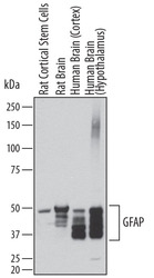
- Experimental details
- Detection of Human and Rat GFAP by Western Blot. Western blot shows lysates of rat cortical stem cells, rat brain tissue, human brain (cortex) tissue, and human brain (hypothalamus) tissue. PVDF membrane was probed with 0.2 µg/mL of Sheep Anti-Human GFAP Antigen Affinity-purified Polyclonal Antibody (Catalog # AF2594) followed by HRP-conjugated Anti-Sheep IgG Secondary Antibody (Catalog # HAF016). Specific bands were detected for GFAP at approximately 35-50 kDa (as indicated). This experiment was conducted under reducing conditions and using Immunoblot Buffer Group 1.
- Submitted by
- R&D Systems (provider)
- Main image
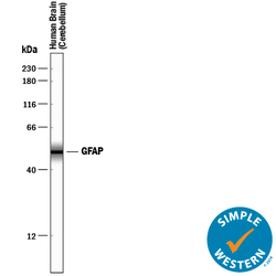
- Experimental details
- Detection of Human GFAP by Simple WesternTM. Simple Western lane view shows lysates of human brain (cerebellum) tissue, loaded at 0.2 mg/mL. A specific band was detected for GFAP at approximately 51 kDa (as indicated) using 0.1 µg/mL of Sheep Anti-Human/Rat GFAP Antigen Affinity-purified Polyclonal Antibody (Catalog # AF2594) followed by 1:50 dilution of HRP-conjugated Anti-Sheep IgG Secondary Antibody (Catalog # HAF016). This experiment was conducted under reducing conditions and using the 12-230 kDa separation system.
- Submitted by
- R&D Systems (provider)
- Main image
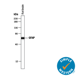
- Experimental details
- Detection of Rat GFAP by Simple WesternTM. Simple Western lane view shows lysates of rat brain tissue, loaded at 0.2 mg/mL. A specific band was detected for GFAP at approximately 55 kDa (as indicated) using 2 µg/mL of Sheep Anti-Human/Rat GFAP Antigen Affinity-purified Polyclonal Antibody (Catalog # AF2594) followed by 1:50 dilution of HRP-conjugated Anti-Sheep IgG Secondary Antibody (Catalog # HAF016). This experiment was conducted under reducing conditions and using the 12-230 kDa separation system.
Supportive validation
- Submitted by
- R&D Systems (provider)
- Main image

- Experimental details
- beta-III Tubulin in Rat Cortical Neurons and GFAP in Rat Astrocytes. beta-III Tubulin was detected in rat cortical neurons using 5 µg/mL Mouse Anti-neuron-specific Mouse beta-III Tubulin Monoclonal (clone TuJ-1) Antibody (Catalog # MAB1195). GFAP was detected in rat astrocytes using 10 µg/mL Sheep Anti-Human GFAP Antigen Affinity-purified Poly-clonal Antibody (Catalog # AF2594). Cells were incubated with primary antibodies for 3 hours at room temperature. Cells were stained for beta-III Tubulin using the Northern-Lights™ 557-conjugated Anti-Mouse IgG Secondary Antibody (red; Catalog # NL007) and for GFAP using the Northern-Lights 493-conjugated Anti-Sheep IgG Secondary Antibody (green; Catalog # NL012). View our protocol for Fluorescent ICC Staining of Cells on Coverslips.
- Submitted by
- R&D Systems (provider)
- Main image
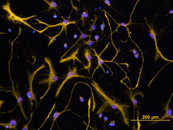
- Experimental details
- GFAP in Rat Cortical Stem Cells. GFAP was detected in immersion fixed 7 days differentiated rat cortical stem cells using Sheep Anti-Human GFAP Antigen Affinity-purified Poly-clonal Antibody (Catalog # AF2594) at 10 µg/mL for 3 hours at room temperature. Cells were stained using the Northern-Lights™ 557-conjugated Anti-Sheep IgG Secondary Antibody (yellow; Catalog # NL010) and counterstained with DAPI (blue). View our protocol for Fluorescent ICC Staining of Cells on Coverslips.
- Submitted by
- R&D Systems (provider)
- Main image
- Experimental details
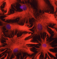
- GFAP in Rat Astrocytes. GFAP was detected in immersion fixed rat astrocytes using 10 µg/mL Sheep Anti-Human GFAP Antigen Affinity-purified Polyclonal Antibody (Catalog # AF2594) for 3 hours at room temperature. Cells were stained with the NorthernLights™ 557-conjugated Anti-Sheep IgG Secondary Antibody (red; Catalog # NL010) and counterstained with DAPI (blue). View our protocol for Fluorescent ICC Staining of Cells on Coverslips.
 Explore
 Validate
 Learn
Learn Western blot