Explore
 Validate
 Learn
Learn Western blot
Antibody data
- Antibody Data
- Antigen structure
- References [6]
- Comments [0]
- Validations
- Western blot [2]
- Immunocytochemistry [1]
Submit
Validation data
Reference
Comment
Report error
- Product number
- MAB2594 - Provider product page

- Provider
- R&D Systems
- Product name
- Human GFAP Antibody
- Antibody type
- Monoclonal
- Description
- Protein A or G purified from hybridoma culture supernatant. Detects human GFAP in direct ELISAs and Western blots.
- Reactivity
- Human
- Host
- Mouse
- Conjugate
- Unconjugated
- Antigen sequence
P14136- Isotype
- IgG
- Antibody clone number
- 273807
- Vial size
- 100 ug
- Concentration
- LYOPH
- Storage
- Use a manual defrost freezer and avoid repeated freeze-thaw cycles. 12 months from date of receipt, -20 to -70 °C as supplied. 1 month, 2 to 8 °C under sterile conditions after reconstitution. 6 months, -20 to -70 °C under sterile conditions after reconstitution.
Submitted references Enhanced sphingosine-1-phosphate receptor 2 expression underlies female CNS autoimmunity susceptibility.
MicroRNA-210 overexpression induces angiogenesis and neurogenesis in the normal adult mouse brain.
Gene expression profile of glioblastoma peritumoral tissue: an ex vivo study.
CD133 positive embryonal rhabdomyosarcoma stem-like cell population is enriched in rhabdospheres.
Presence of pluripotent CD133+ cells correlates with malignancy of gliomas.
Development of a culture system that supports adult microglial cell proliferation and maintenance in the resting state.
Cruz-Orengo L, Daniels BP, Dorsey D, Basak SA, Grajales-Reyes JG, McCandless EE, Piccio L, Schmidt RE, Cross AH, Crosby SD, Klein RS
The Journal of clinical investigation 2014 Jun;124(6):2571-84
The Journal of clinical investigation 2014 Jun;124(6):2571-84
MicroRNA-210 overexpression induces angiogenesis and neurogenesis in the normal adult mouse brain.
Zeng L, He X, Wang Y, Tang Y, Zheng C, Cai H, Liu J, Wang Y, Fu Y, Yang GY
Gene therapy 2014 Jan;21(1):37-43
Gene therapy 2014 Jan;21(1):37-43
Gene expression profile of glioblastoma peritumoral tissue: an ex vivo study.
Mangiola A, Saulnier N, De Bonis P, Orteschi D, Sica G, Lama G, Pettorini BL, Sabatino G, Zollino M, Lauriola L, Colabianchi A, Proietti G, Kovacs G, Maira G, Anile C
PloS one 2013;8(3):e57145
PloS one 2013;8(3):e57145
CD133 positive embryonal rhabdomyosarcoma stem-like cell population is enriched in rhabdospheres.
Walter D, Satheesha S, Albrecht P, Bornhauser BC, D'Alessandro V, Oesch SM, Rehrauer H, Leuschner I, Koscielniak E, Gengler C, Moch H, Bernasconi M, Niggli FK, Schäfer BW, CWS Study Group.
PloS one 2011;6(5):e19506
PloS one 2011;6(5):e19506
Presence of pluripotent CD133+ cells correlates with malignancy of gliomas.
Thon N, Damianoff K, Hegermann J, Grau S, Krebs B, Schnell O, Tonn JC, Goldbrunner R
Molecular and cellular neurosciences 2010 Jan;43(1):51-9
Molecular and cellular neurosciences 2010 Jan;43(1):51-9
Development of a culture system that supports adult microglial cell proliferation and maintenance in the resting state.
Ponomarev ED, Novikova M, Maresz K, Shriver LP, Dittel BN
Journal of immunological methods 2005 May;300(1-2):32-46
Journal of immunological methods 2005 May;300(1-2):32-46
No comments: Submit comment
Supportive validation
- Submitted by
- R&D Systems (provider)
- Main image
- Experimental details
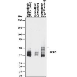
- Detection of Human GFAP by Western Blot. Western blot shows lysates of human brain (motor cortex) tissue, human brain (cerebellum) tissue, and human brain (hypothalamus) tissue. PVDF membrane was probed with 1 µg/mL of Mouse Anti-Human GFAP Monoclonal Antibody (Catalog # MAB2594) followed by HRP-conjugated Anti-Mouse IgG Secondary Antibody (Catalog # HAF018). Specific bands were detected for GFAP at approximately 35-50 kDa (as indicated). This experiment was conducted under reducing conditions and using Immunoblot Buffer Group 1.

- Submitted by
- R&D Systems (provider)
- Main image
- Experimental details
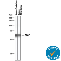
- Detection of Human GFAP by Simple WesternTM. Simple Western lane view shows lysates of human brain (cerebellum) tissue and human brain (motor cortex) tissue, loaded at 0.2 mg/mL. A specific band was detected for GFAP at approximately 51-52 kDa (as indicated) using 50 µg/mL of Mouse Anti-Human GFAP Monoclonal Antibody (Catalog # MAB2594) . This experiment was conducted under reducing conditions and using the 12-230 kDa separation system.

Supportive validation
- Submitted by
- R&D Systems (provider)
- Main image

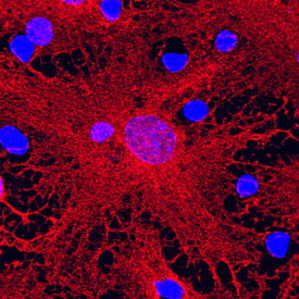
- Experimental details
- GFAP in Rat Cortical Stem Cells. GFAP was detected in immersion fixed differentiated rat cortical stem cells using Mouse Anti-Human GFAP Monoclonal Antibody (Catalog # MAB2594) at 10 µg/mL for 3 hours at room temperature. Cells were stained using the NorthernLights™ 557-conjugated Anti-Mouse IgG Secondary Antibody (red; Catalog # NL007) and counterstained with DAPI (blue). Specific staining was localized to cytoplasm. View our protocol for Fluorescent ICC Staining of Stem Cells on Coverslips.