Explore
 Validate
 Learn
Learn Western blot
 ELISA
ELISAAntibody data
- Antibody Data
- Antigen structure
- References [0]
- Comments [0]
- Validations
- Western blot [4]
Submit
Validation data
Reference
Comment
Report error
- Product number
- PA1-26402 - Provider product page

- Provider
- Invitrogen Antibodies
- Product name
- Myeloperoxidase Polyclonal Antibody
- Antibody type
- Polyclonal
- Antigen
- Other
- Description
- PA1-26402 detects Myeloperoxidase from human samples.
- Reactivity
- Human
- Host
- Rabbit
- Isotype
- IgG
- Vial size
- 100 µL
- Concentration
- 85 mg/mL
- Storage
- Store at 4°C short term. For long term storage, store at -20°C, avoiding freeze/thaw cycles.
No comments: Submit comment
Supportive validation
- Submitted by
- Invitrogen Antibodies (provider)
- Main image

- Experimental details
- Anti-Myeloperoxidase (Human Leukocytes) detects multiple MPO subunits and chain combinations by Western blot. Polyclonal rabbit-anti-Myeloperoxidase (Product # PA1-26402) was used at a 1:5000 dilution to detect 1.0 µg of human myeloperoxidase. This antibody detects a multiple bands corresponding to 53 kDa and 15 kDa polypeptides and chain combinations forming 68 kDa and 106 kDa proteins. The staining of the 68 kDa band is so intense that is over saturates the signal detection. A 4-20% gradient gel was used to separate the protein by SDS-PAGE. The protein was transferred to nitrocellulose using standard methods. After blocking the membrane was probed with the primary antibody for 2 h at room temperature followed by washes and reaction with a 1:5000 dilution of infrared 800 conjugated Goat-anti-Rabbit IgG (H&L) for 30 min at room temperature.

- Submitted by
- Invitrogen Antibodies (provider)
- Main image

- Experimental details
- Anti-Myeloperoxidase [Human Leukocytes] detects multiple MPO subunits and chain combinations by western blot. Polyclonal rabbit-anti-Myeloperoxidase Myeloperoxidase Polyclonal Antibody (Product # PA1-26402) was used at a 1:5,000 dilution to detect 1.0 µg of human myeloperoxidase. This antibody detects a multiple bands corresponding to 53 kDa and 15 kDa polypeptides and chain combinations forming 68 kDa and 106 kDa proteins. The staining of the 68 kDa band is so intense that is over saturates the signal detection. A 4-20% gradient gel was used to separate the protein by SDS-PAGE. The protein was transferred to nitrocellulose using standard methods. After blocking the membrane was probed with the primary antibody for 2 h at room temperature followed by washes and reaction with a 1:5,000 dilution of IRDye™800 conjugated Goat anti Rabbit IgG [H&L] for 30 min at room temperature.

- Submitted by
- Invitrogen Antibodies (provider)
- Main image

- Experimental details
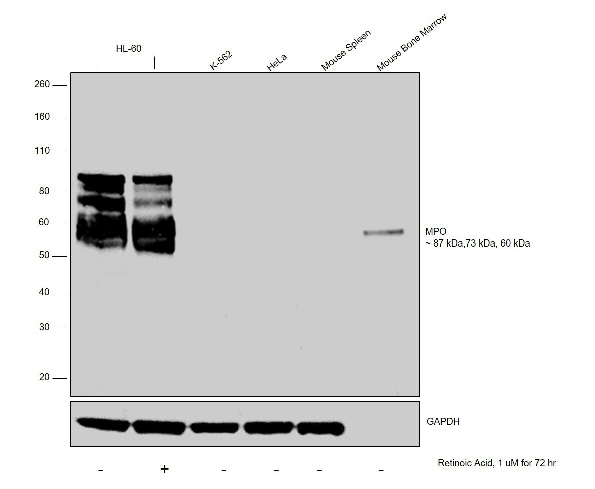
- Western blot was performed using Anti-Myeloperoxidase Rabbit Polyclonal Antibody (Product # PA1-26402) and 87 kDa, 73 kDa and 60 kDa bands corresponding to MPO was observed across cell lines and tissues tested except K-562, HeLa and Mouse Spleen. Membrane enriched extracts (30 µg lysate) of HL-60 (Lane 1), HL-60 treated with Retinoic Acid (1uM for 72 hr) (Lane 2), K-562 (Lane 3), HeLa (Lane 4), tissue extracts (30 ug lysate) of Mouse Spleen (Lane 5) and Mouse Bone Marrow (Lane 6) were electrophoresed using NuPAGE™ 4-12% Bis-Tris Protein Gel (Product # NP0322BOX). Resolved proteins were then transferred onto a nitrocellulose membrane (Product # IB23001) by iBlot® 2 Dry Blotting System (Product # IB21001). The blot was probed with the primary antibody (1:1500 dilution) and detected by chemiluminescence with Goat anti-Rabbit IgG (H+L), Superclonal™ Recombinant Secondary Antibody, HRP (Product # A27036, 1:4000 dilution) using the iBright FL 1000 (Product # A32752). Chemiluminescent detection was performed using Novex® ECL Chemiluminescent Substrate Reagent Kit (Product # WP20005).