 Explore
 Validate
 Learn
Learn Western blot
Antibody data
- Antibody Data
- Antigen structure
- References [4]
- Comments [0]
- Validations
- Western blot [1]
- Immunocytochemistry [2]
- Immunohistochemistry [1]
Submit
Validation data
Reference
Comment
Report error
- Product number
- MAB3174 - Provider product page

- Provider
- R&D Systems
- Product name
- Human Myeloperoxidase/MPO Antibody
- Antibody type
- Monoclonal
- Description
- Protein A or G purified from hybridoma culture supernatant. Detects human Myeloperoxidase/MPO in Western blots. No cross-reactivity with recombinant human Eosinophil Peroxidase is observed.
- Reactivity
- Human
- Host
- Mouse
- Conjugate
- Unconjugated
- Antigen sequence
P05164- Isotype
- IgG
- Antibody clone number
- 392105
- Vial size
- 100 ug
- Concentration
- LYOPH
- Storage
- Use a manual defrost freezer and avoid repeated freeze-thaw cycles. 12 months from date of receipt, -20 to -70 °C as supplied. 1 month, 2 to 8 °C under sterile conditions after reconstitution. 6 months, -20 to -70 °C under sterile conditions after reconstitution.
Submitted references The Traditional Chinese Medicine MLC901 inhibits inflammation processes after focal cerebral ischemia.
Antimicrobial cathelicidin peptide LL‑37 induces NET formation and suppresses the inflammatory response in a mouse septic model.
Myocardial Structural and Biological Anomalies Induced by High Fat Diet in Psammomys obesus Gerbils.
The Ancient Immunoglobulin Domains of Peroxidasin Are Required to Form Sulfilimine Cross-links in Collagen IV.
Widmann C, Gandin C, Petit-Paitel A, Lazdunski M, Heurteaux C
Scientific reports 2018 Dec 24;8(1):18062
Scientific reports 2018 Dec 24;8(1):18062
Antimicrobial cathelicidin peptide LL‑37 induces NET formation and suppresses the inflammatory response in a mouse septic model.
Hosoda H, Nakamura K, Hu Z, Tamura H, Reich J, Kuwahara-Arai K, Iba T, Tabe Y, Nagaoaka I
Molecular medicine reports 2017 Oct;16(4):5618-5626
Molecular medicine reports 2017 Oct;16(4):5618-5626
Myocardial Structural and Biological Anomalies Induced by High Fat Diet in Psammomys obesus Gerbils.
Sahraoui A, Dewachter C, de Medina G, Naeije R, Aouichat Bouguerra S, Dewachter L
PloS one 2016;11(2):e0148117
PloS one 2016;11(2):e0148117
The Ancient Immunoglobulin Domains of Peroxidasin Are Required to Form Sulfilimine Cross-links in Collagen IV.
Ero-Tolliver IA, Hudson BG, Bhave G
The Journal of biological chemistry 2015 Aug 28;290(35):21741-8
The Journal of biological chemistry 2015 Aug 28;290(35):21741-8
No comments: Submit comment
Supportive validation
- Submitted by
- R&D Systems (provider)
- Main image
- Experimental details
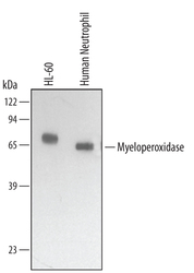
- Detection of Human Myeloperoxidase/MPO by Western Blot. Western blot shows lysates of HL-60 human acute promyelocytic leukemia cell line and human neutrophil. PVDF membrane was probed with 1 µg/mL of Mouse Anti-Human Myeloperoxidase/MPO Monoclonal Antibody (Catalog # MAB3174) followed by HRP-conjugated Anti-Mouse IgG Secondary Antibody (Catalog # HAF007). A specific band was detected for Myeloperoxidase/MPO at approximately 65 kDa (as indicated). This experiment was conducted under reducing conditions and using Immunoblot Buffer Group 2.

Supportive validation
- Submitted by
- R&D Systems (provider)
- Main image
- Experimental details
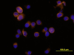
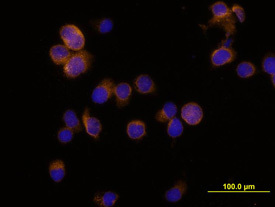
- Myeloperoxidase/MPO in HL-60 Human Cell Line. Myeloperoxidase/MPO was detected in immersion fixed HL-60 human acute promyelocytic leukemia cell line using Mouse Anti-Human Myeloperoxidase/MPO Mono-clonal Antibody (Catalog # MAB3174) at 10 µg/mL for 3 hours at room temperature. Cells were stained using the NorthernLights™ 557-conju-gated Anti-Mouse IgG Secondary Antibody (yellow; Catalog # NL007) and counterstained with DAPI (blue). View our protocol for Fluorescent ICC Staining of Non-adherent Cells.
- Submitted by
- R&D Systems (provider)
- Main image

- Experimental details
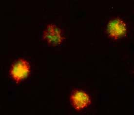
- Myeloperoxidase/MPO in MOLT-4 Human Cell Line. Myeloperoxidase/ MPO was detected in immersion fixed MOLT-4 human acute lymphoblastic leukemia cell line using 8 µg/mL Mouse Anti-Human Myeloperoxidase/ MPO Monoclonal Antibody (Catalog # MAB3174) for 3 hours at room temperature. Cells were stained (red) and counter-stained (green). View our protocol for Fluorescent ICC Staining of Cells on Coverslips.
Supportive validation
- Submitted by
- R&D Systems (provider)
- Main image
- Experimental details
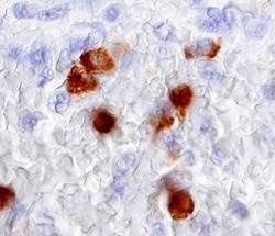
- Myeloperoxidase/MPO in Human Spleen. Myeloperoxidase/MPO was detected in immersion fixed paraffin-embedded sections of human spleen using 15 µg/mL Mouse Anti-Human Myeloperoxidase/MPO Monoclonal Antibody (Catalog # MAB3174) overnight at 4 °C. Before incubation with the primary antibody tissue was subjected to heat-induced epitope retrieval using Antigen Retrieval Reagent-Basic (Catalog # CTS013). Tissue was stained with the Anti-Mouse HRP-DAB Cell & Tissue Staining Kit (brown; Catalog # CTS002) and counterstained with hematoxylin (blue). View our protocol for Chromogenic IHC Staining of Paraffin-embedded Tissue Sections.
