Explore
 Validate
 Learn
Learn Western blot
Antibody data
- Antibody Data
- Antigen structure
- References [0]
- Comments [0]
- Validations
- Western blot [3]
- Immunohistochemistry [10]
Submit
Validation data
Reference
Comment
Report error
- Product number
- AMAb90710 - Provider product page

- Provider
- Atlas Antibodies
- Proper citation
- Atlas Antibodies Cat#AMAb90710, RRID:AB_2665641
- Product name
- Anti-P4HA2
- Antibody type
- Monoclonal
- Reactivity
- Human
- Host
- Mouse
- Conjugate
- Unconjugated
- Antigen sequence
LKEYILVEEAKLSKIKSWANKMEALTSKSAADAEG
YLAHPVNAYKLVKRLNTDWPALEDLVLQDSAAGFI
ANLSVQRQFFPTDEDEIGAAKALMRLQDTYRLDPG
TISRGELPGTKYQAMLSVDDCFGM- Epitope
- Binds to an epitope located within the peptide sequence NTDWPALEDLVLQDS as determined by overlapping synthetic peptides.
- Isotype
- IgG
- Antibody clone number
- CL0351
- Vial size
- 100 µl
- Storage
- Store at +4°C for short term storage. Long time storage is recommended at -20°C.
No comments: Submit comment
Enhanced validation
- Submitted by
- Atlas Antibodies (provider)
- Enhanced method
- Genetic validation
- Main image

- Experimental details
- Western blot analysis in U-251MG cells transfected with control siRNA, target specific siRNA probe #1 and #2, using Anti-P4HA2 antibody. Remaining relative intensity is presented. Loading control: Anti-GAPDH.

- Submitted by
- Atlas Antibodies (provider)
- Main image
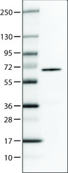
- Experimental details
- Lane 1: Marker [kDa]Lane 2: Human cell line U-251 MG

- Submitted by
- Atlas Antibodies (provider)
- Main image
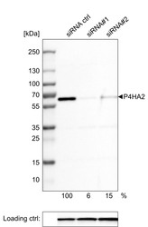
- Experimental details
- Western blot analysis of extracts from U-251 cells, transfected with: control siRNA, target specific siRNA probe #1, target specific siRNA probe #2, using Anti-P4HA2 monoclonal antibody. Downregulation of antibody signal confirms target specificity. Remaining % intensity, relative control lane, is indicated. Anti-GAPDH monoclonal antibody was used as loading control.

Enhanced validation
Supportive validation
- Submitted by
- Atlas Antibodies (provider)
- Enhanced method
- Orthogonal validation
- Main image
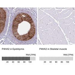
- Experimental details
- Immunohistochemistry analysis in human epididymis and skeletal muscle tissues using AMAb90710 antibody. Corresponding P4HA2 RNA-seq data are presented for the same tissues.
- Sample type
- HUMAN

Supportive validation
- Submitted by
- Atlas Antibodies (provider)
- Main image

- Experimental details
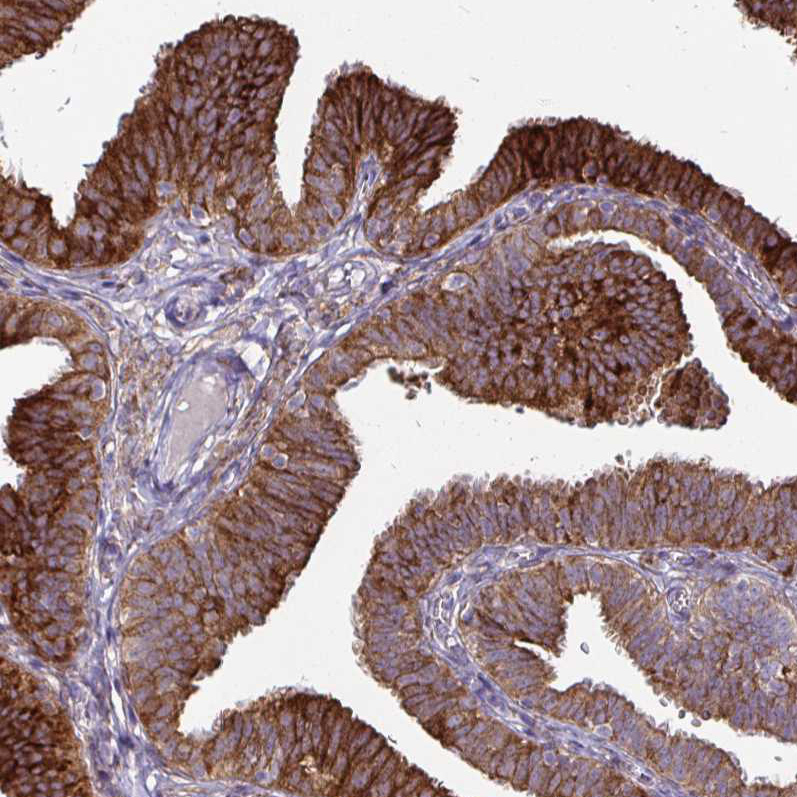
- Immunohistochemical staining of human endometrium shows strong cytoplasmic immunoreactivity in glandular epithelium, as well as in underlying connective tissue.
- Submitted by
- Atlas Antibodies (provider)
- Main image
- Experimental details
- Immunohistochemical staining of human testis shows moderate to strong positivity in seminiferous duct cells.
- Submitted by
- Atlas Antibodies (provider)
- Main image

- Experimental details
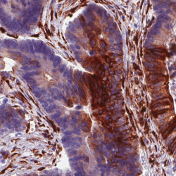
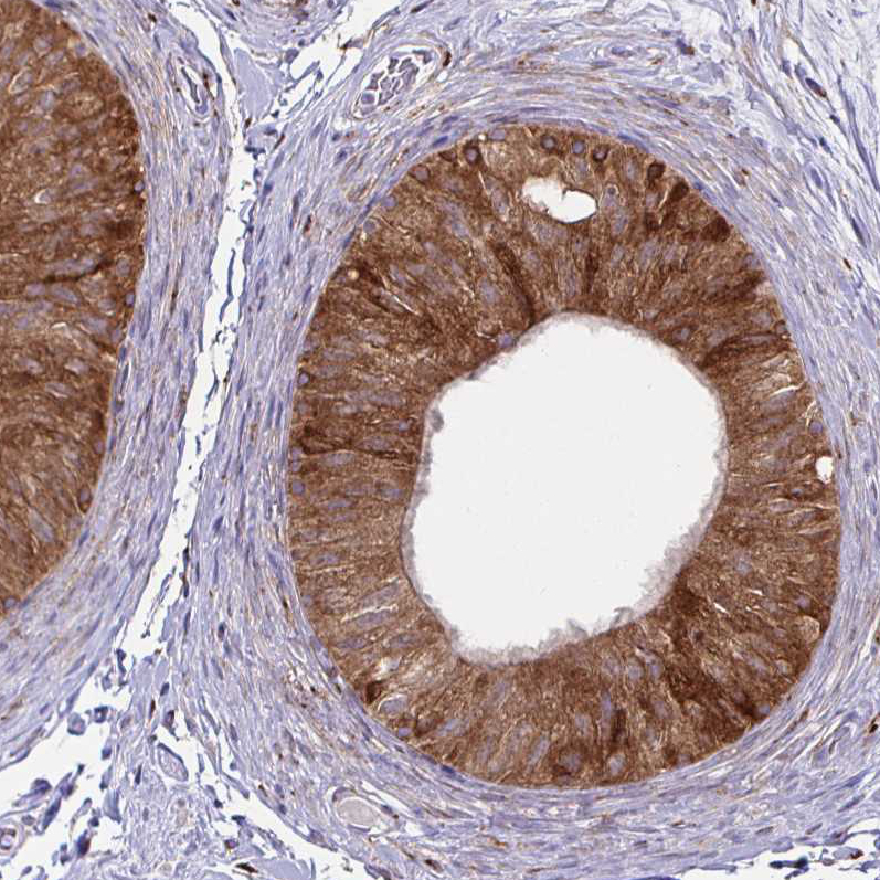
- Immunohistochemical staining of human duodenum shows moderate immunoreactivity in glandular cells and underlying connective tissue.

- Submitted by
- Atlas Antibodies (provider)
- Main image
- Experimental details
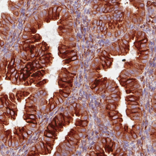
- Immunohistochemical staining of human colorectal cancer shows moderate to strong positivity in the cancer cells and underlying connective tissue.

- Submitted by
- Atlas Antibodies (provider)
- Main image
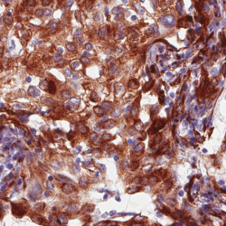
- Experimental details
- Immunohistochemical staining of human lung cancer (lung adenocarcinoma) shows strong immunorectivity in the tumour cells.

- Submitted by
- Atlas Antibodies (provider)
- Main image

- Experimental details
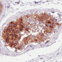
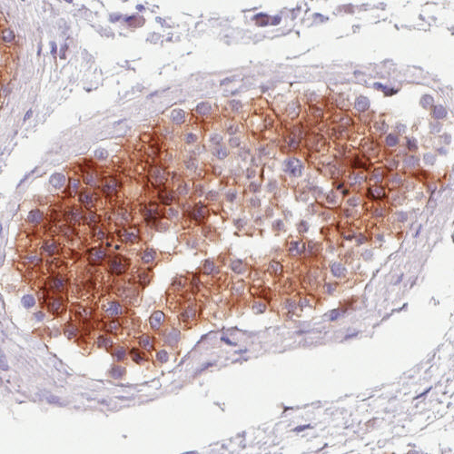
- Immunohistochemical staining of human epididymis shows moderate to strong cytoplasmic positivity in glandular cells.
- Submitted by
- Atlas Antibodies (provider)
- Main image

- Experimental details
- Immunohistochemical staining of human fallopian tube shows moderate to strong cytoplasmic positivity in glandular cells.
- Submitted by
- Atlas Antibodies (provider)
- Main image
- Experimental details
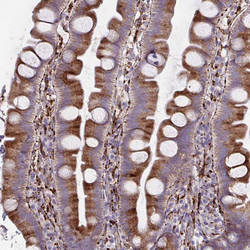
- Immunohistochemical staining of human small intestine shows moderate cytoplasmic positivity in glandular cells.

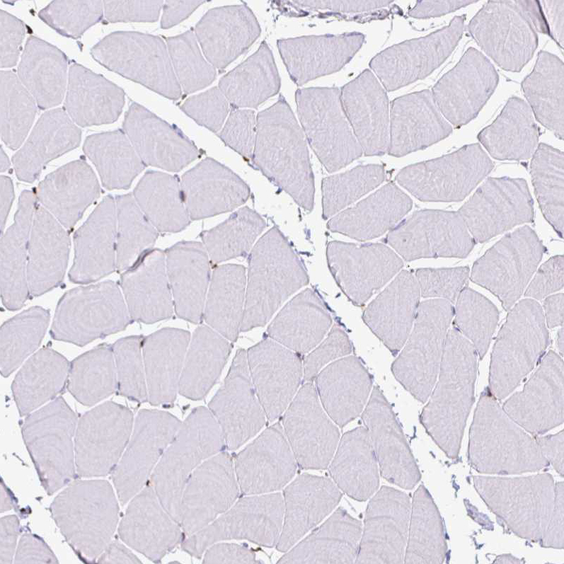
- Submitted by
- Atlas Antibodies (provider)
- Main image

- Experimental details
- Immunohistochemical staining of human skeletal muscle shows no positivity in striated muscle fibers as expected.