Explore
 Validate
 Learn
Learn Western blot
Antibody data
- Antibody Data
- Antigen structure
- References [2]
- Comments [0]
- Validations
- Western blot [2]
- Immunohistochemistry [2]
Submit
Validation data
Reference
Comment
Report error
- Product number
- GTX101553 - Provider product page

- Provider
- GeneTex
- Proper citation
- GeneTex Cat#GTX101553, RRID:AB_1240765
- Product name
- NSE antibody [N1C1]
- Antibody type
- Polyclonal
- Reactivity
- Human, Mouse, Rat
- Host
- Rabbit
Submitted references Continued 26S proteasome dysfunction in mouse brain cortical neurons impairs autophagy and the Keap1-Nrf2 oxidative defence pathway.
REST reduction is essential for hypoxia-induced neuroendocrine differentiation of prostate cancer cells by activating autophagy signaling.
Ugun-Klusek A, Tatham MH, Elkharaz J, Constantin-Teodosiu D, Lawler K, Mohamed H, Paine SM, Anderson G, John Mayer R, Lowe J, Ellen Billett E, Bedford L
Cell death & disease 2017 Jan 5;8(1):e2531
Cell death & disease 2017 Jan 5;8(1):e2531
REST reduction is essential for hypoxia-induced neuroendocrine differentiation of prostate cancer cells by activating autophagy signaling.
Lin TP, Chang YT, Lee SY, Campbell M, Wang TC, Shen SH, Chung HJ, Chang YH, Chiu AW, Pan CC, Lin CH, Chu CY, Kung HJ, Cheng CY, Chang PC
Oncotarget 2016 May 3;7(18):26137-51
Oncotarget 2016 May 3;7(18):26137-51
No comments: Submit comment
Supportive validation
- Submitted by
- GeneTex (provider)
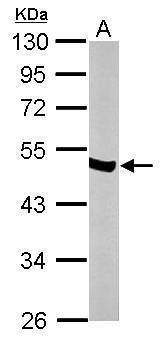
- Main image

- Experimental details
- Sample (50 ?g of whole cell lysate) A: mouse brain 10% SDS PAGE GTX101553 diluted at 1:10000 The HRP-conjugated anti-rabbit IgG antibody (GTX213110-01) was used to detect the primary antibody.
- Submitted by
- GeneTex (provider)
- Main image
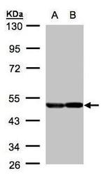
- Experimental details
- Sample(30 ?g of whole cell lysate)A:A431(GTX27909)B:H129910% SDS PAGEGTX101553 diluted at 1:2000The HRP-conjugated anti-rabbit IgG antibody (GTX213110-01) was used to detect the primary antibody.
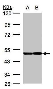
Supportive validation
- Submitted by
- GeneTex (provider)
- Main image
- Experimental details
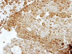
- Immunohistochemical analysis of paraffin-embedded PC14 xenograft, using ENO2(GTX101553) antibody at 1:100 dilution.

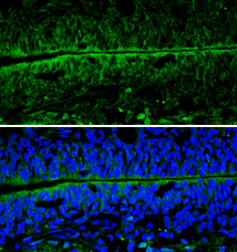
- Submitted by
- GeneTex (provider)
- Main image
- Experimental details
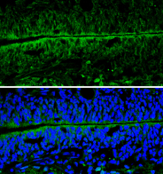
- NSE antibody [N1C1] detects NSE protein expression by immunohistochemical analysis.Sample: Frozen sectioned E13.5 Rat brain. Green: NSE protein stained by NSE antibody [N1C1] (GTX101553) diluted at 1:250.Blue: Fluoroshield with DAPI (GTX30920).