 Explore
 Validate
 Learn
Learn Western blot
 Immunocytochemistry
Antibody data
- Antibody Data
- Antigen structure
- References [3]
- Comments [0]
- Validations
- Western blot [2]
- Immunocytochemistry [1]
- Immunohistochemistry [8]
Submit
Validation data
Reference
Comment
Report error
- Product number
- HPA000834 - Provider product page

- Provider
- Atlas Antibodies
- Proper citation
- Atlas Antibodies Cat#HPA000834, RRID:AB_1078977
- Product name
- Anti-G6PD
- Antibody type
- Polyclonal
- Reactivity
- Human
- Host
- Rabbit
- Conjugate
- Unconjugated
- Antigen sequence
FQGDAFHQSDTHIFIIMGASGDLAKKKIYPTIWWL
FRDGLLPENTFIVGYARSRLTVADIRKQSEPFFKA
TPEEKLKLEDFFARNSYVAGQYDDAASYQRLNSHM
NALH- Isotype
- IgG
- Vial size
- 100 µl
- Storage
- Store at +4°C for short term storage. Long time storage is recommended at -20°C.
Submitted references Global variability in gene expression and alternative splicing is modulated by mitochondrial content
TAp73 enhances the pentose phosphate pathway and supports cell proliferation.
Variance decomposition of protein profiles from antibody arrays using a longitudinal twin model.
Guantes R, Rastrojo A, Neves R, Lima A, Aguado B, Iborra F
Genome Research 2015 May;25(5):633-644
Genome Research 2015 May;25(5):633-644
TAp73 enhances the pentose phosphate pathway and supports cell proliferation.
Du W, Jiang P, Mancuso A, Stonestrom A, Brewer MD, Minn AJ, Mak TW, Wu M, Yang X
Nature cell biology 2013 Aug;15(8):991-1000
Nature cell biology 2013 Aug;15(8):991-1000
Variance decomposition of protein profiles from antibody arrays using a longitudinal twin model.
Kato BS, Nicholson G, Neiman M, Rantalainen M, Holmes CC, Barrett A, Uhlén M, Nilsson P, Spector TD, Schwenk JM
Proteome science 2011 Nov 17;9:73
Proteome science 2011 Nov 17;9:73
No comments: Submit comment
Enhanced validation
Enhanced validation
- Submitted by
- Atlas Antibodies (provider)
- Enhanced method
- Orthogonal validation
- Main image
- Experimental details
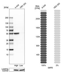
- Western blot analysis in human cell lines A-549 and HEK293 using Anti-G6PD antibody. Corresponding G6PD RNA-seq data are presented for the same cell lines. Loading control: Anti-HSP90B1.

Enhanced validation
- Submitted by
- Atlas Antibodies (provider)
- Enhanced method
- Independent antibody validation
- Main image
- Experimental details
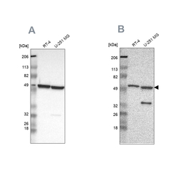
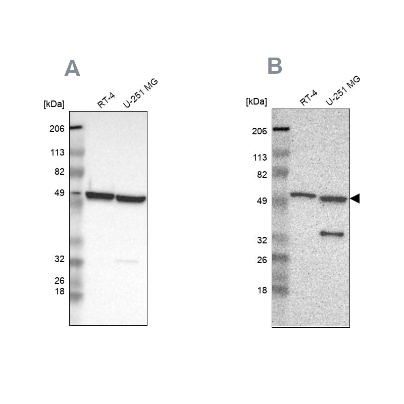
- Western blot analysis using Anti-G6PD antibody HPA000834 (A) shows similar pattern to independent antibody HPA000247 (B).
Supportive validation
- Submitted by
- Atlas Antibodies (provider)
- Main image
- Experimental details
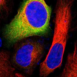
- Immunofluorescent staining of human cell line U-2 OS shows positivity in cytoplasm.
- Sample type
- HUMAN

Enhanced validation
Supportive validation
- Submitted by
- Atlas Antibodies (provider)
- Enhanced method
- Orthogonal validation
- Main image
- Experimental details
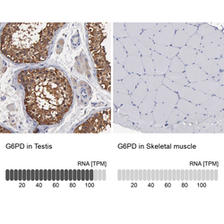
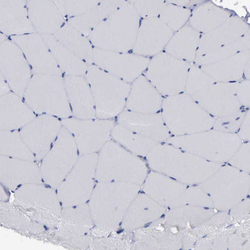
- Immunohistochemistry analysis in human testis and skeletal muscle tissues using HPA000834 antibody. Corresponding G6PD RNA-seq data are presented for the same tissues.
- Sample type
- HUMAN

Supportive validation
- Submitted by
- Atlas Antibodies (provider)
- Main image
- Experimental details
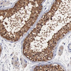
- Immunohistochemical staining of human testis shows high expression.
- Sample type
- HUMAN

- Submitted by
- Atlas Antibodies (provider)
- Main image
- Experimental details
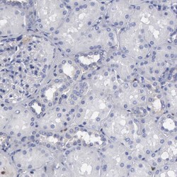
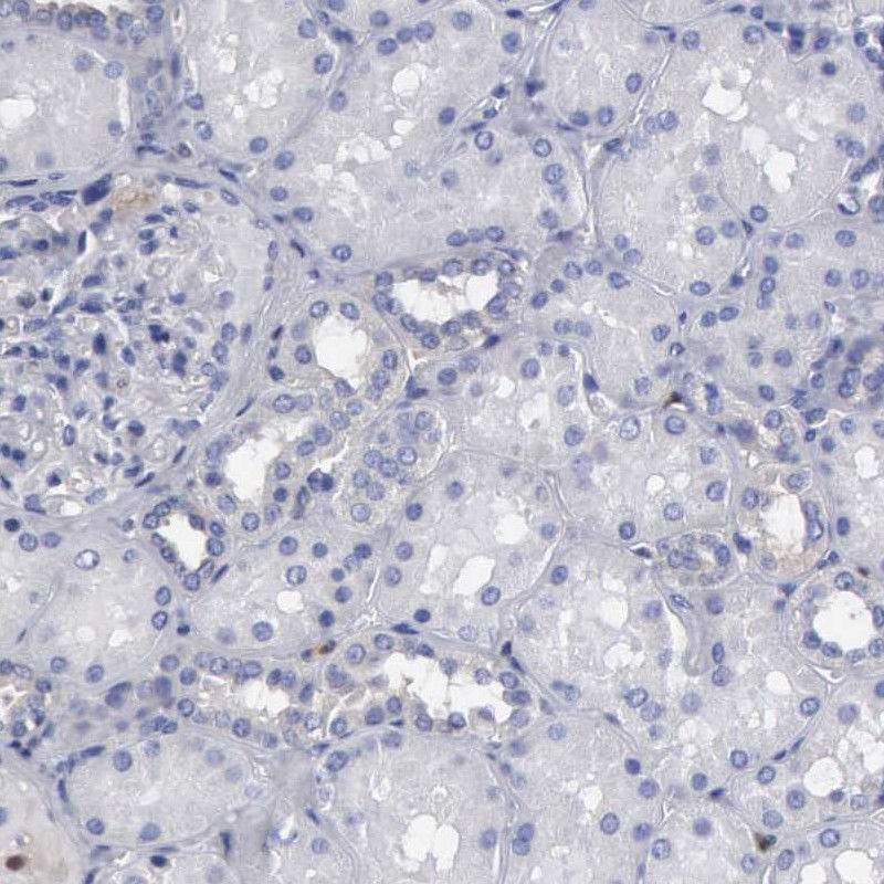
- Immunohistochemical staining of human kidney shows low expression as expected.
- Sample type
- HUMAN
- Submitted by
- Atlas Antibodies (provider)
- Main image
- Experimental details
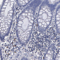
- Immunohistochemical staining of human colon shows moderate cytoplasmic positivity in a subset of lymphoid cells.
- Sample type
- HUMAN

- Submitted by
- Atlas Antibodies (provider)
- Main image

- Experimental details
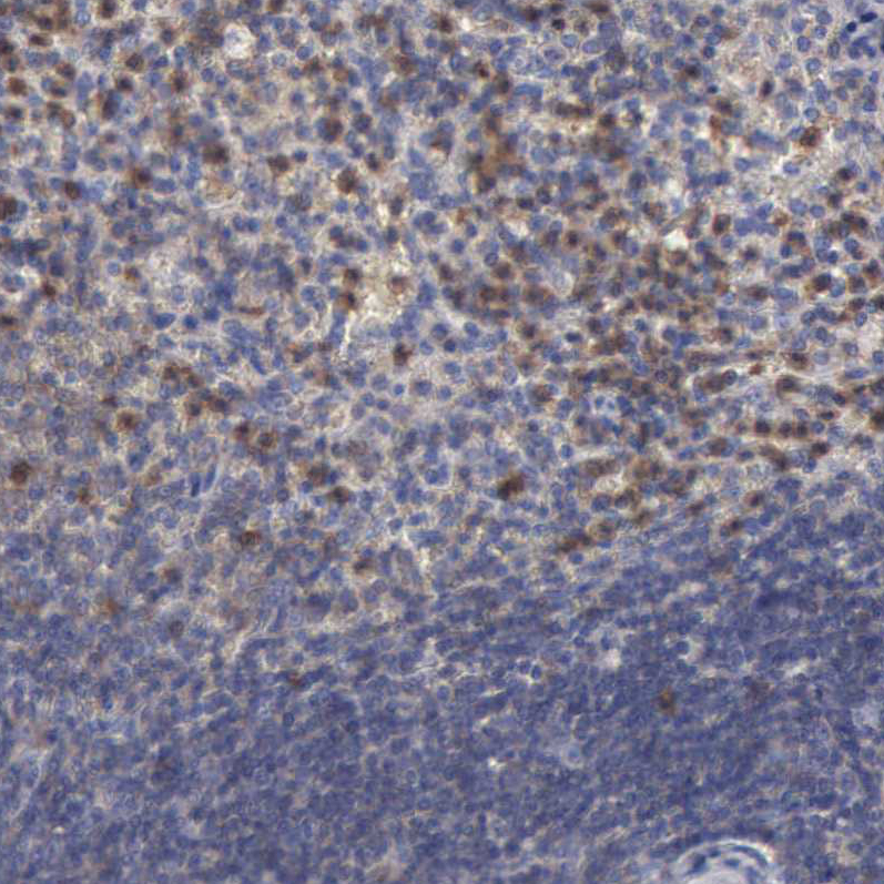
- Immunohistochemical staining of human spleen shows moderate cytoplasmic positivity in lymphoid cells.
- Sample type
- HUMAN
- Submitted by
- Atlas Antibodies (provider)
- Main image
- Experimental details
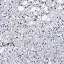
- Immunohistochemical staining of human liver shows moderate to strong cytoplasmic positivity in Kupffer cells.
- Sample type
- HUMAN

- Submitted by
- Atlas Antibodies (provider)
- Main image
- Experimental details
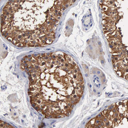
- Immunohistochemical staining of human testis shows strong cytoplasmic positivity in cells in seminiferous ducts.
- Sample type
- HUMAN

- Submitted by
- Atlas Antibodies (provider)
- Main image
- Experimental details
- Immunohistochemical staining of human skeletal muscle shows no cytoplasmic positivity in myocytes as expected.
- Sample type
- HUMAN
