Explore
 Validate
 Learn
Learn Western blot
 ELISA
ELISAAntibody data
- Antibody Data
- Antigen structure
- References [1]
- Comments [0]
- Validations
- Western blot [6]
- Immunocytochemistry [1]
- Immunohistochemistry [2]
- Flow cytometry [1]
Submit
Validation data
Reference
Comment
Report error
- Product number
- MA5-15918 - Provider product page

- Provider
- Invitrogen Antibodies
- Product name
- G6PD Monoclonal Antibody (5E12)
- Antibody type
- Monoclonal
- Antigen
- Purifed from natural sources
- Reactivity
- Human
- Host
- Mouse
- Isotype
- IgG
- Antibody clone number
- 5E12
- Vial size
- 100 µL
- Concentration
- Conc. Not Determined
- Storage
- Store at 4°C short term. For long term storage, store at -20°C, avoiding freeze/thaw cycles.
Submitted references Placental growth factor regulates the pentose phosphate pathway and antioxidant defense systems in human retinal endothelial cells.
Saddala MS, Lennikov A, Huang H
Journal of proteomics 2020 Apr 15;217:103682
Journal of proteomics 2020 Apr 15;217:103682
No comments: Submit comment
Supportive validation
- Submitted by
- Invitrogen Antibodies (provider)
- Main image

- Experimental details
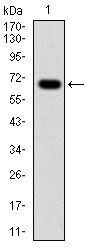
- Western blot analysis of G6PD using a G6PD monoclonal antibody (Product # MA5-15918) against a human G6PD (AA: 275-515) recombinant protein.

- Submitted by
- Invitrogen Antibodies (provider)
- Main image

- Experimental details
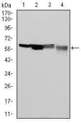
- Western blot analysis of G6PD using G6PD monoclonal antibody (Product # MA5-15918) in HeLa (1), MCF-7 (2), Jurkat (3) and K562 (4) cell lysate.
- Submitted by
- Invitrogen Antibodies (provider)
- Main image
- Experimental details
- Western blot analysis of G6PD using a G6PD monoclonal antibody (Product # MA5-15918) against a human G6PD (AA: 275-515) recombinant protein.

- Submitted by
- Invitrogen Antibodies (provider)
- Main image
- Experimental details
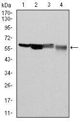
- Western blot analysis of G6PD using G6PD monoclonal antibody (Product # MA5-15918) in HeLa (1), MCF-7 (2), Jurkat (3) and K562 (4) cell lysate.

- Submitted by
- Invitrogen Antibodies (provider)
- Main image
- Experimental details
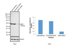
- Knockdown of G6PD was achieved by transfecting HeLa cells with G6PD specific siRNAs (Silencer® select Product # s5447). Western blot analysis (Fig. a) was performed whole cell extracts from the HeLa knockdown cells (lane 3), non-specific scrambled siRNA transfected cells (lane 2) and untransfected cells (lane 1). The blots were probed with G6PD Monoclonal Antibody (Product # MA5-15918, 1:1000) and Goat anti-Mouse IgG (H+L) Superclonal™ Recombinant Secondary Antibody, HRP (Product # A28177, 1:4000 dilution). Densitometric analysis of this western blot is shown in histogram (Fig. b). Decrease in signal upon siRNA mediated knock down confirms that antibody is specific to G6PD.

- Submitted by
- Invitrogen Antibodies (provider)
- Main image
- Experimental details
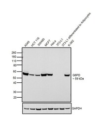
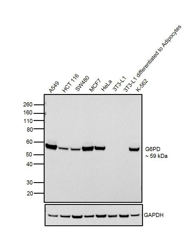
- Western blot was performed using Anti-G6PD Mouse Monoclonal Antibody (Product # MA5-15918) and a 59 kDa band corresponding to G6PD was observed across cell lines tested. Whole cell extracts (30 µg lysate) of A549 (Lane 1), HCT 116 (Lane 2), SW480 (Lane 2), MCF7 (Lane 4), HeLa (Lane 5), 3T3-L1 (Lane 6), 3T3-L1 differentiated to Adipocytes (Lane 7) and K-562 (Lane 8) were electrophoresed using NuPAGE™ 4-12% Bis-Tris Protein Gel (Product # NP0322BOX). Resolved proteins were then transferred onto a nitrocellulose membrane (Product # IB23001) by iBlot® 2 Dry Blotting System (Product # IB21001). The blot was probed with the primary antibody (1:1000 dilution) and detected by chemiluminescence with Goat anti-Mouse IgG (H+L), Superclonal™ Recombinant Secondary Antibody, HRP (Product # A28177, 1:4000 dilution) using the iBright FL 1000 (Product # A32752). Chemiluminescent detection was performed using Novex® ECL Chemiluminescent Substrate Reagent Kit (Product # WP20005). This product does not show reactivity to mouse G6PD, as no band was observed in mouse 3T3-L1 cell line when differentiated to adipocytes.
Supportive validation
- Submitted by
- Invitrogen Antibodies (provider)
- Main image
- Experimental details
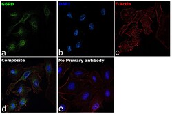
- Immunofluorescence analysis of G6PD was performed using A549 cells. The cells were fixed with 4% paraformaldehyde for 10 minutes, permeabilized with 0.1% Triton™ X-100 for 15 minutes, and blocked with 2% BSA for 1 hour at room temperature. The cells were labeled with G6PD Monoclonal Antibody (5E12) (Product # MA5-15918) at 1:250 dilution in 0.1% BSA and incubated overnight at 4 degree and then labeled with Goat anti-Mouse IgG (H+L) Superclonal™ Recombinant Secondary Antibody, Alexa Fluor® 488 conjugate (Product # A28175) at a dilution of 1:2000 for 45 minutes at room temperature (Panel a: green). Nuclei (Panel b: blue) were stained with ProLong™ Diamond Antifade Mountant with DAPI (Product # P36962). F-actin (Panel c: red) was stained with Rhodamine Phalloidin (Product # R415, 1:300). Panel d represents the composite image showing Nuclear and Cytoplasmic localization of G6PD. Panel e represents control cells with no primary antibody to assess background. The images were captured at 60X magnification.

Supportive validation
- Submitted by
- Invitrogen Antibodies (provider)
- Main image
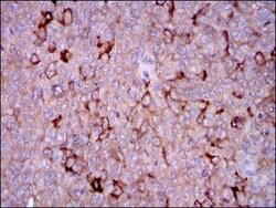
- Experimental details
- Immunohistochemical analysis of paraffin-embedded ovarian cancer tissues using G6PD monoclonal antibody (Product # MA5-15918) followed with DAB staining.
- Submitted by
- Invitrogen Antibodies (provider)
- Main image

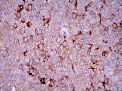
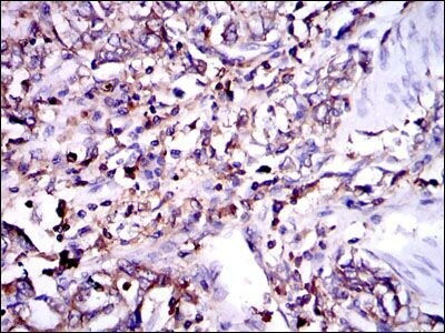
- Experimental details
- Immunohistochemical analysis of paraffin-embedded stomach cancer tissues using G6PD monoclonal antibody (Product # MA5-15918) followed with DAB staining.
Supportive validation
- Submitted by
- Invitrogen Antibodies (provider)
- Main image

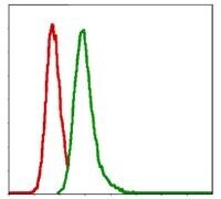
- Experimental details
- Flow cytometric analysis of MCF-7 cells using G6PD monoclonal antibody (Product # MA5-15918) (green) and negative control (red).