Explore
 Validate
 Learn
Learn Western blot
Antibody data
- Antibody Data
- Antigen structure
- References [1]
- Comments [0]
- Validations
- Western blot [4]
- ELISA [1]
- Immunohistochemistry [1]
Submit
Validation data
Reference
Comment
Report error
- Product number
- PA5-18734 - Provider product page

- Provider
- Invitrogen Antibodies
- Product name
- G6PD Polyclonal Antibody
- Antibody type
- Polyclonal
- Antigen
- Synthetic peptide
- Description
- This antibody is predicted to react with canine, mouse and rat based on sequence homology. This antibody is tested in Peptide ELISA: antibody detection limit dilution 32,000.
- Reactivity
- Human
- Host
- Goat
- Isotype
- IgG
- Vial size
- 100 µg
- Concentration
- 0.5 mg/mL
- Storage
- -20° C, Avoid Freeze/Thaw Cycles
Submitted references Downregulation of glucose‑6‑phosphate dehydrogenase by microRNA‑1 inhibits the growth of pituitary tumor cells.
He C, Yang J, Ding J, Li S, Wu H, Xiong Y, Zhou F, Jiang Y, Teng L, Yang J
Oncology reports 2018 Dec;40(6):3533-3542
Oncology reports 2018 Dec;40(6):3533-3542
No comments: Submit comment
Supportive validation
- Submitted by
- Invitrogen Antibodies (provider)
- Main image
- Experimental details
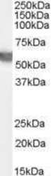
- Western Blot staining of Human Testes lysate using Product # PA5-18734 at a concentration of 0.03 µg/mL, the primary antibody incubation was 1 hour and the detection method was chemiluminescence.

- Submitted by
- Invitrogen Antibodies (provider)
- Main image

- Experimental details
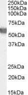
- Western Blot staining of Human Testes lysate using Product # PA5-18734 at a concentration of 0.03 µg/mL, the primary antibody incubation was 1 hour and the detection method was chemiluminescence.
- Submitted by
- Invitrogen Antibodies (provider)
- Main image

- Experimental details
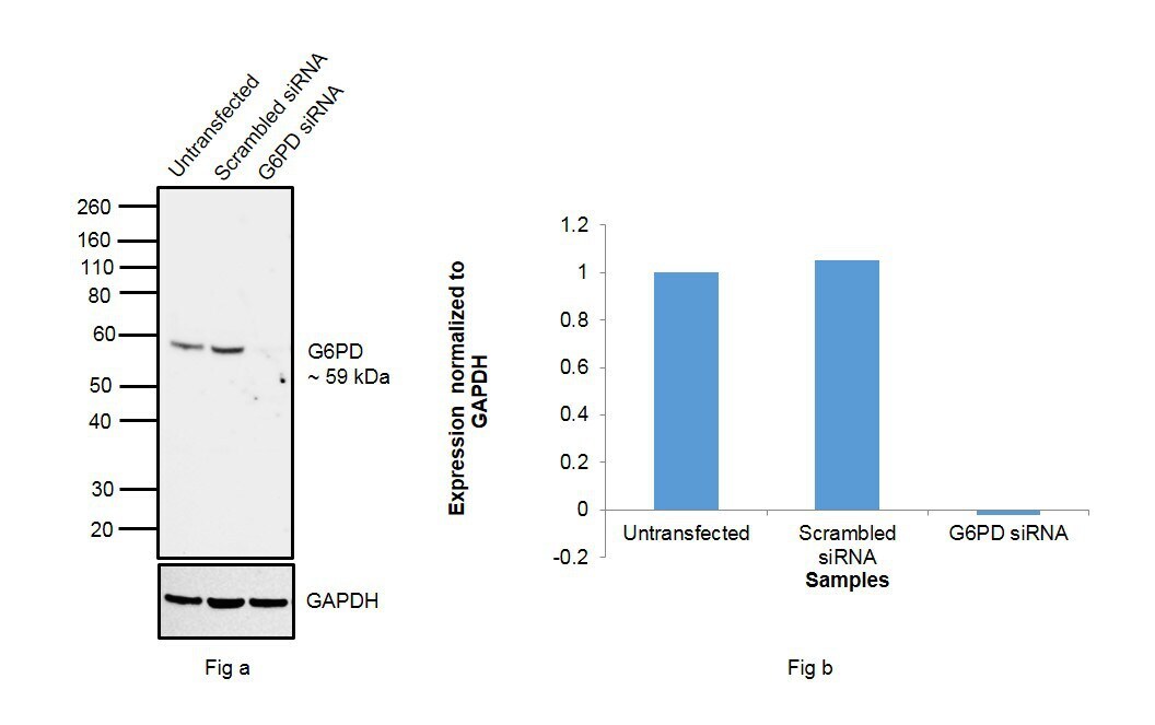
- Knockdown of G6PD was achieved by transfecting HeLa cells with G6PD specific siRNAs (Silencer® select Product # s5447). Western blot analysis (Fig. a) was performed whole cell extracts from the HeLa knockdown cells (lane 3), non-specific scrambled siRNA transfected cells (lane 2) and untransfected cells (lane 1). The blots were probed with G6PD Polyclonal Antibody (Product # PA5-18734, 1:500) and Rabbit anti-Goat IgG (H+L) Superclonal™ Recombinant Secondary Antibody, HRP (Product # A27014, 1:4000 dilution). Densitometric analysis of this western blot is shown in histogram (Fig. b). Decrease in signal upon siRNA mediated knock down confirms that antibody is specific to G6PD.
- Submitted by
- Invitrogen Antibodies (provider)
- Main image
- Experimental details
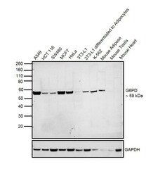
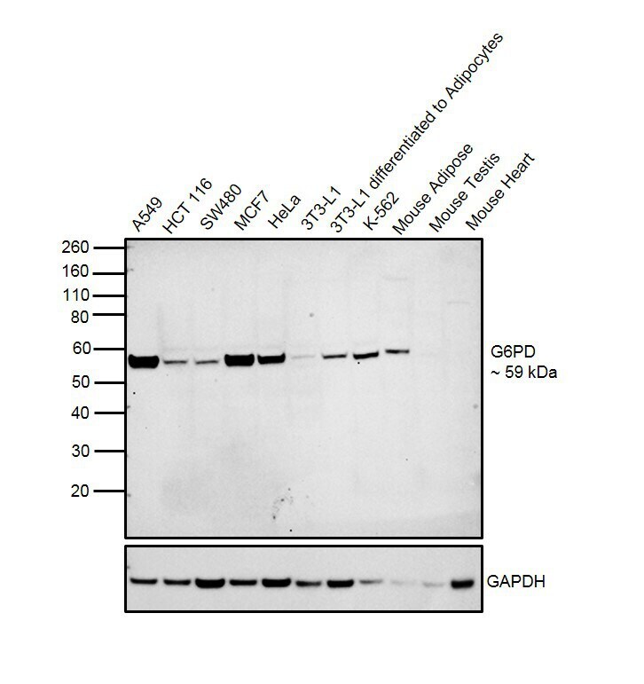
- Western blot was performed using Anti-G6PD Goat Polyclonal Antibody (Product # PA5-18734) and a 59 kDa band corresponding to G6PD was observed across cell lines tested and tissues tested except Mouse Testis and Mouse Heart. Whole cell extracts (30 µg lysate) of A549 (Lane 1), HCT 116 (Lane 2), SW480 (Lane 2), MCF7 (Lane 4), HeLa (Lane 5), 3T3-L1 (Lane 6), 3T3-L1 differentiated to Adipocytes (Lane 7), K-562 (Lane 8), Mouse Adipose (Lane 9), Mouse Testis (Lane 10) and Mouse Heart (Lane 11) were electrophoresed using NuPAGE™ 4-12% Bis-Tris Protein Gel (Product # NP0322BOX). Resolved proteins were then transferred onto a nitrocellulose membrane (Product # IB23001) by iBlot® 2 Dry Blotting System (Product # IB21001). The blot was probed with the primary antibody (1:500 dilution) and detected by chemiluminescence with Rabbit anti-Goat IgG (H+L), Superclonal™ Recombinant Secondary Antibody, HRP (Product # A27014, 1:4000 dilution) using the iBright FL 1000 (Product # A32752). Chemiluminescent detection was performed using Novex® ECL Chemiluminescent Substrate Reagent Kit (Product # WP20005).
Supportive validation
- Submitted by
- Invitrogen Antibodies (provider)
- Main image
- Experimental details
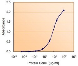
- ELISA of G6PD using two G6PD antibodies. The reporter antibody (Product # PA5-18734) was used at a concentration of 5 µg/ml and the reporter antibody was used at a concentration of 5 µg/ml.

Supportive validation
- Submitted by
- Invitrogen Antibodies (provider)
- Main image

- Experimental details
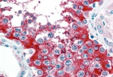
- Immunohistochemical staining of paraffin embedded of Human Testis using Product # PA5-18734 at a concentration of 2.5 µg/mL. The tissue was processed by steamed antigen retrieval with citrate buffer pH 6 and stained with AP.