Antibody data
- Antibody Data
- Antigen structure
- References [3]
- Comments [0]
- Validations
- Western blot [2]
- Immunocytochemistry [1]
- Immunohistochemistry [1]
- Flow cytometry [1]
Submit
Validation data
Reference
Comment
Report error
- Product number
- MAB3696 - Provider product page

- Provider
- R&D Systems
- Product name
- Human c-Myc Antibody
- Antibody type
- Monoclonal
- Description
- Protein A or G purified from hybridoma culture supernatant. Detects endogenous human c-Myc and c-Myc tagged proteins in Western blots.
- Reactivity
- Human
- Host
- Mouse
- Conjugate
- Unconjugated
- Antigen sequence
P01106- Isotype
- IgG
- Antibody clone number
- 9.00E+10
- Vial size
- 100 ug
- Concentration
- LYOPH
- Storage
- Use a manual defrost freezer and avoid repeated freeze-thaw cycles. 12 months from date of receipt, -20 to -70 °C as supplied. 1 month, 2 to 8 °C under sterile conditions after reconstitution. 6 months, -20 to -70 °C under sterile conditions after reconstitution.
Submitted references Critical relevance of genomic gains of PRL-3/EGFR/c-myc pathway genes in liver metastasis of colorectal cancer.
The binding of the bone morphogenetic protein antagonist gremlin to kidney heparan sulfate: Such binding is not essential for BMP antagonism.
U1 Adaptors Suppress the KRAS-MYC Oncogenic Axis in Human Pancreatic Cancer Xenografts.
Tanaka T, Kaida T, Yokoi K, Ishii S, Nishizawa N, Kawamata H, Katoh H, Sato T, Nakamura T, Watanabe M, Yamashita K
Oncology letters 2019 Jan;17(1):1257-1266
Oncology letters 2019 Jan;17(1):1257-1266
The binding of the bone morphogenetic protein antagonist gremlin to kidney heparan sulfate: Such binding is not essential for BMP antagonism.
Tatsinkam AJ, Rune N, Smith J, Norman JT, Mulloy B, Rider CC
The international journal of biochemistry & cell biology 2017 Feb;83:39-46
The international journal of biochemistry & cell biology 2017 Feb;83:39-46
U1 Adaptors Suppress the KRAS-MYC Oncogenic Axis in Human Pancreatic Cancer Xenografts.
Tsang AT, Dudgeon C, Yi L, Yu X, Goraczniak R, Donohue K, Kogan S, Brenneman MA, Ho ES, Gunderson SI, Carpizo DR
Molecular cancer therapeutics 2017 Aug;16(8):1445-1455
Molecular cancer therapeutics 2017 Aug;16(8):1445-1455
No comments: Submit comment
Supportive validation
- Submitted by
- R&D Systems (provider)
- Main image
- Experimental details
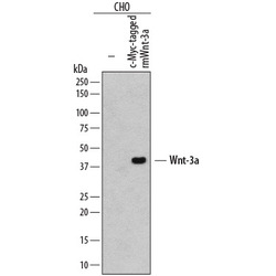
- Detection of c-Myc-tagged Protein by Western Blot. Western blot shows lysates of CHO Chinese hamster ovary cell line either mock transfected (-) or transfected with c-Myc-tagged recombinant mouse Wnt-3a. PVDF membrane was probed with 2 µg/mL of Mouse Anti-Human c-Myc Monoclonal Antibody (Catalog # MAB3696) followed by HRP-conjugated Anti-Mouse IgG Secondary Antibody (Catalog # HAF007). A specific band was detected for c-Myc-tagged recombinant mouse Wnt-3a at approximately 41 kDa (as indicated). This experiment was conducted under reducing conditions and using Immunoblot Buffer Group 1.
- Submitted by
- R&D Systems (provider)
- Main image
- Experimental details
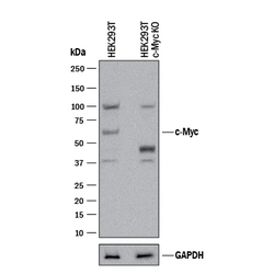
- Western Blot Shows Human c-Myc Specificity by Using Knockout Cell Line. Western blot shows lysates of HEK293T human embryonic kidney parental cell line and c-Myc knockout HEK293T cell line (KO). PVDF membrane was probed with 2 µg/mL of Mouse Anti-Human c-Myc Monoclonal Antibody (Catalog # MAB3696) followed by HRP-conjugated Anti-Mouse IgG Secondary Antibody (Catalog # HAF018). A specific band was detected for c-Myc at approximately 52 kDa (as indicated) in the parental HEK293T cell line, but is not detectable in knockout HEK293T cell line. GAPDH (Catalog # MAB5718) is shown as a loading control. This experiment was conducted under reducing conditions and using Immunoblot Buffer Group 1.
Supportive validation
- Submitted by
- R&D Systems (provider)
- Main image
- Experimental details
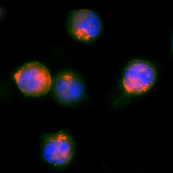
- c-Myc in HEK293 Human Cell Line Transfected with c-Myc-tagged Serotonin Receptor. c-Myc was detected in immersion fixed HEK293 human embryonic kidney cell line transfected with c-Myc-tagged Serotonin Receptor using Mouse Anti-Human c-Myc Monoclonal Antibody (Catalog # MAB3696) at 25 µg/mL for 3 hours at room temperature. Cells were stained using the NorthernLights™ 557-conjugated Anti-Mouse IgG Secondary Antibody (red; Catalog # NL007) and counterstained with DAPI (blue). Specific staining was localized to nuclei. View our protocol for Fluorescent ICC Staining of Cells on Coverslips.
Supportive validation
- Submitted by
- R&D Systems (provider)
- Main image
- Experimental details
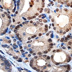
- c-Myc in Human Prostate. c-Myc was detected in immersion fixed paraffin-embedded sections of human prostate using Mouse Anti-Human c-Myc Monoclonal Antibody (Catalog # MAB3696) at 3 µg/mL overnight at 4 °C. Tissue was stained using the Anti-Mouse HRP-DAB Cell & Tissue Staining Kit (brown; Catalog # CTS002) and counterstained with hematoxylin (blue). Specific staining was localized to nuclei of epithelial cells. View our protocol for Chromogenic IHC Staining of Paraffin-embedded Tissue Sections.
Supportive validation
- Submitted by
- R&D Systems (provider)
- Main image
- Experimental details
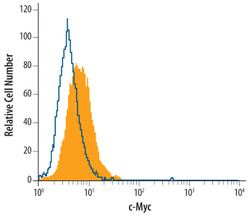
- Detection of c-Myc in Jurkat Human Cell Line by Flow Cytometry. Jurkat human acute T cell leukemia cell line was stained with Mouse Anti-Human c-Myc Mono-clonal Antibody (Catalog # MAB3696, filled histogram) or isotype control antibody (Catalog # MAB002, open histogram), followed by Phycoerythrin-conjugated Anti-Mouse IgG Secondary Antibody (Catalog # F0102B). To facilitate intracellular staining, cells were fixed with paraformaldehyde and permeabilized with methanol.
 Explore
 Validate
 Learn
Learn Western blot
 Immunoprecipitation