 Explore
 Validate
 Learn
Learn Western blot
 Immunoprecipitation
Antibody data
- Antibody Data
- Antigen structure
- References [7]
- Comments [0]
- Validations
- Western blot [7]
- ELISA [2]
- Immunocytochemistry [2]
- Immunohistochemistry [1]
- Other assay [6]
Submit
Validation data
Reference
Comment
Report error
- Product number
- PA5-29578 - Provider product page

- Provider
- Invitrogen Antibodies
- Product name
- Fibronectin Polyclonal Antibody
- Antibody type
- Polyclonal
- Antigen
- Recombinant protein fragment
- Description
- Recommended positive controls: HepG2, HepG2(10 mM Nicotinamide and 0.4 mM Trichostatin A treatment for 48 hr), HeLa mock and shFN1, mouse plasma. Predicted reactivity: Mouse (92%), Rat (92%), Chicken (88%), Bovine (92%). Store product as a concentrated solution. Centrifuge briefly prior to opening the vial.
- Reactivity
- Human, Mouse, Rat
- Host
- Rabbit
- Isotype
- IgG
- Vial size
- 100 µL
- Concentration
- 0.48 mg/mL
- Storage
- Store at 4°C short term. For long term storage, store at -20°C, avoiding freeze/thaw cycles.
Submitted references Paclitaxel Induces Epidermal Molecular Changes and Produces Subclinical Alterations in the Skin of Gynecological Cancer Patients.
Human Mesenchymal Stromal Cell-Derived Exosomes Promote In Vitro Wound Healing by Modulating the Biological Properties of Skin Keratinocytes and Fibroblasts and Stimulating Angiogenesis.
A step toward engineering thick tissues: Distributing microfibers within 3D printed frames.
1,25 Dihydroxyvitamin D3 Enhances the Antifibroid Effects of Ulipristal Acetate in Human Uterine Fibroids.
Diet-induced hepatic steatosis abrogates cell-surface LDLR by inducing de novo PCSK9 expression in mice.
Morphological and Molecular Changes in Juvenile Normal Human Fibroblasts Exposed to Simulated Microgravity.
In vitro assessment of a novel, hypothermically stored amniotic membrane for use in a chronic wound environment.
Montero P, Pérez-Leal M, Pérez-Fidalgo JA, Sanz C, Estornut C, Roger I, Milara J, Cervantes A, Cortijo J
Cancers 2022 Feb 23;14(5)
Cancers 2022 Feb 23;14(5)
Human Mesenchymal Stromal Cell-Derived Exosomes Promote In Vitro Wound Healing by Modulating the Biological Properties of Skin Keratinocytes and Fibroblasts and Stimulating Angiogenesis.
Tutuianu R, Rosca AM, Iacomi DM, Simionescu M, Titorencu I
International journal of molecular sciences 2021 Jun 9;22(12)
International journal of molecular sciences 2021 Jun 9;22(12)
A step toward engineering thick tissues: Distributing microfibers within 3D printed frames.
Molde J, Steele JAM, Pastino AK, Mahat A, Murthy NS, Kohn J
Journal of biomedical materials research. Part A 2020 Mar;108(3):581-591
Journal of biomedical materials research. Part A 2020 Mar;108(3):581-591
1,25 Dihydroxyvitamin D3 Enhances the Antifibroid Effects of Ulipristal Acetate in Human Uterine Fibroids.
Ali M, Shahin SM, Sabri NA, Al-Hendy A, Yang Q
Reproductive sciences (Thousand Oaks, Calif.) 2019 Jun;26(6):812-828
Reproductive sciences (Thousand Oaks, Calif.) 2019 Jun;26(6):812-828
Diet-induced hepatic steatosis abrogates cell-surface LDLR by inducing de novo PCSK9 expression in mice.
Lebeau PF, Byun JH, Platko K, MacDonald ME, Poon SV, Faiyaz M, Seidah NG, Austin RC
The Journal of biological chemistry 2019 Jun 7;294(23):9037-9047
The Journal of biological chemistry 2019 Jun 7;294(23):9037-9047
Morphological and Molecular Changes in Juvenile Normal Human Fibroblasts Exposed to Simulated Microgravity.
Buken C, Sahana J, Corydon TJ, Melnik D, Bauer J, Wehland M, Krüger M, Balk S, Abuagela N, Infanger M, Grimm D
Scientific reports 2019 Aug 15;9(1):11882
Scientific reports 2019 Aug 15;9(1):11882
In vitro assessment of a novel, hypothermically stored amniotic membrane for use in a chronic wound environment.
McQuilling JP, Vines JB, Mowry KC
International wound journal 2017 Dec;14(6):993-1005
International wound journal 2017 Dec;14(6):993-1005
No comments: Submit comment
Supportive validation
- Submitted by
- Invitrogen Antibodies (provider)
- Main image

- Experimental details
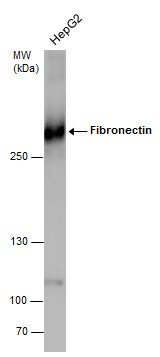
- Western blot analysis of Fibronectin using Whole cell extracts (30 µg). Samples were loaded onto a 5% SDS-PAGE gel and probed with a Fibronectin polyclonal antibody (Product # PA5-29578) at a dilution of 1:1000.

- Submitted by
- Invitrogen Antibodies (provider)
- Main image

- Experimental details
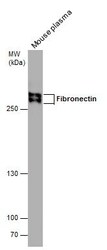
- Western blot analysis of Fibronectin in Mouse tissue extracts (50 µg). Samples was separated by 5% SDS-PAGE and the membrane was probed with Fibronectin Polyclonal antibody (Product # PA5-29578) at a dilution of 1:1000.

- Submitted by
- Invitrogen Antibodies (provider)
- Main image

- Experimental details
- Western Blot analysis of Fibronectin was performed by separating 30 µg of whole cell extracts by 5% SDS-PAGE. Proteins were transferred to a membrane and probed with a Fibronectin Polyclonal Antibody (Product # PA5-29578) at a dilution of 1:1000.
- Submitted by
- Invitrogen Antibodies (provider)
- Main image

- Experimental details
- Knockout of Fibronectin was achieved by CRISPR-Cas9 genome editing using LentiArray™ Lentiviral sgRNA (Product # A32042, Assay ID CRISPR616644_LV) and LentiArray Cas9 Lentivirus (Product # A32064). Western blot analysis of Fibronectin was performed by loading 30 µg of Hep G2 Wild Type (Lane 1), Hep G2 Wild Type treated with 1X PTI for 4hrs (Lane 2),Hep G2 Cas9 (Lane 3), Hep G2 Cas9 treated with 1X PTI for 4hrs (Lane 4), Hep G2 Fibronectin KO (Lane 5) and Hep G2 Fibronectin KO treated with 1X PTI for 4hrs (Lane 6) whole cell extracts. The samples were electrophoresed using NuPAGE™ 3-8% Tris-Acetate Protein Gel (Product # EA0378BOX). Resolved proteins were then transferred onto a nitrocellulose membrane (Product # IB23001) by iBlot® 2 Dry Blotting System (Product # IB21001). The blot was probed with Anti-Fibronectin Polyclonal Antibody (Product # PA5-29578, 0.1 µg/mL dilution) and Goat anti-Rabbit IgG (H+L) Superclonal™ Recombinant Secondary Antibody, HRP (Product # A27036, 1:5,000 dilution) using the iBright FL 1000 (Product # A32752). Chemiluminescent detection was performed using Novex® ECL Chemiluminescent Substrate Reagent Kit (Product # WP20005). Loss of signal upon CRISPR mediated knockout (KO) using the LentiArray™ CRISPR product line confirms that antibody is specific to Fibronectin.

- Submitted by
- Invitrogen Antibodies (provider)
- Main image
- Experimental details
- Fibronectin Polyclonal Antibody [N1N2] detects Fibronectin protein by western blot analysis. Mouse tissue extracts (50 µg) was separated by 5% SDS-PAGE, and the membrane was blotted with Fibronectin Polyclonal Antibody [N1N2] Fibronectin Polyclonal Antibody (Product # PA5-29578) diluted at 1:1,000. The HRP-conjugated anti-rabbit IgG antibody was used to detect the primary antibody.

- Submitted by
- Invitrogen Antibodies (provider)
- Main image

- Experimental details
- Western Blot analysis of Fibronectin was performed by separating 30 µg of untreated (–) and treated (+) HepG2 whole cell extracts by 5% SDS-PAGE. Proteins were transferred to a membrane and probed with a Fibronectin Polyclonal Antibody (Product # PA5-29578) at a dilution of 1:2000.

- Submitted by
- Invitrogen Antibodies (provider)
- Main image

- Experimental details
- Western blot was performed using Anti-Fibronectin Polyclonal Antibody (Product # PA5-29578) and a ~260 kDa band corresponding to Fibronectin was observed across cell lines tested. Whole cell extracts (30 µg lysate) of Hep G2 (Lane 1), Hep G2 treated with 1X PTI for 4h (Lane 2), Hep G2 treated with 10 µm FLI-06 for 4h (Lane 3), HEL 92.1.7 (Lane 4), HEL 92.1.7 treated with 1X PTI for 4h (Lane 5), HEL 92.1.7 treated with 10 µm FLI-06 for 4h (Lane 6) were electrophoresed using NuPAGE™ 3-8% Tris-Acetate Protein Gel (Product # EA0378BOX). Resolved proteins were then transferred onto a Nitrocellulose membrane (Product # IB23001) by iBlot® 2 Dry Blotting System (Product # IB21001). The blot was probed with the primary antibody (0.14 µg/mL) and detected by chemiluminescence with Goat anti-Rabbit IgG (H+L) Superclonal™ Recombinant Secondary Antibody, HRP (Product # A27036, 1:4,000) using the iBright FL 1000 (Product # A32752). Chemiluminescent detection was performed using Novex® ECL Chemiluminescent Substrate Reagent Kit (Product # WP20005). Another isoform of Fibronectin (*) was seen in the cell lines around 120 kDa. The antibody showed enhanced signal in positive cell line Hep G2 upon treatment with secretion blockers as compared to the negative cell line HEL 92.1.7 treated with the same blockers.

Supportive validation
- Submitted by
- Invitrogen Antibodies (provider)
- Main image

- Experimental details
- Competitive ELISA detection of human fibronectin in plasma using Fibronectin Polyclonal Antibody (Product # PA5-29578). Recombinant or purified native Fibronectin protein was coated to an ELISA plate at concentration of 10 µg/mL. Human plasma was applied as the competitors and was serial diluted at 1, 20, 200, 2,000, 20,000, 200,000X dilutions. The primary antibody concentration was diluted to 14 ng/mL. Rabbit IgG antibody (HRP) was diluted at 1:2,000 and used to detect the primary antibody.

- Submitted by
- Invitrogen Antibodies (provider)
- Main image

- Experimental details
- Indirect ELISA analysis was performed by coating plate with 100 µL of recombinant Fibronectin protein at concentration of 10 µg/mL. The coated protein is detected with Fibronectin Polyclonal Antibody (Product # PA5-29578) at rangeing from 0.5 to 140 ng/mL.

Supportive validation
- Submitted by
- Invitrogen Antibodies (provider)
- Main image
- Experimental details
- Immunofluorescent analysis of Fibronectin in paraformaldehyde-fixed HeLa cells using a Fibronectin polyclonal antibody (Product # PA5-29578) at a 1:200 dilution.

- Submitted by
- Invitrogen Antibodies (provider)
- Main image

- Experimental details
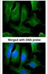
- Immunofluorescence analysis of Fibronectin was performed using 70% confluent log phase Hep G2 cells. The cells were fixed with 4% paraformaldehyde for 10 minutes, permeabilized with 0.1% Triton™ X-100 for 15 minutes, and blocked with 2% BSA for 45 minutes at room temperature. The cells were labeled with Fibronectin Polyclonal Antibody (Product # PA5-29578) at 1:200 in 0.1% BSA, incubated at 4 degree celsius overnight and then labeled with Donkey anti-Rabbit IgG (H+L) Highly Cross-Adsorbed Secondary Antibody, Alexa Fluor Plus 488 (Product # A32790), (1:2000), for 45 minutes at room temperature (Panel a: Green). Nuclei (Panel b:Blue) were stained with ProLong™ Diamond Antifade Mountant with DAPI (Product # P36962). F-actin (Panel c: Red) was stained with Rhodamine Phalloidin (Product # R415, 1:300). Panel d represents the merged image showing cytosolic localization. Panel e represents untreated Hep G2 cells with no expression. Panel f represents control cells with no primary antibody to assess background The images were captured at 60X magnification.

Supportive validation
- Submitted by
- Invitrogen Antibodies (provider)
- Main image

- Experimental details
- Fibronectin Polyclonal Antibody detects FN1 protein at cytosol on human hepatoma by immunohistochemical analysis. Sample: Paraffin-embedded hepatoma. Fibronectin Polyclonal Antibody (Product # PA5-29578) dilution: 1:500. Antigen Retrieval: EDTA based buffer, pH 8.0, 15 min.

Supportive validation
- Submitted by
- Invitrogen Antibodies (provider)
- Main image

- Experimental details
- Fibronectin Polyclonal Antibody immunoprecipitates Fibronectin protein in IP experiments. IP Sample: HeLa whole cell lysate/extract A : 30 µg whole cell lysate/extract of Fibronectin protein expressing HeLa cells B : Control with 3 µg of pre-immune rabbit IgG C : Immunoprecipitation of Fibronectin by 3 µg of Fibronectin Polyclonal Antibody (Product # PA5-29578) 5% SDS-PAGE The immunoprecipitated Fibronectin protein was detected by Fibronectin Polyclonal Antibody (Product # PA5-29578) diluted at 1:1,000. Anti-rabbit IgG (HRP) was used as a secondary reagent.

- Submitted by
- Invitrogen Antibodies (provider)
- Main image

- Experimental details
- Figure 7 Qualitative evaluation of fibroblast interaction with hypothermically stored amniotic membrane was completed using immunohistochemical stains. Representative images were taken of collagen I (A and B), collagen II (C and D) and fibronectin (E and F). Black arrows point to fibronectin deposited throughout the matrix. IWJ-12748-FIG-0007-c

- Submitted by
- Invitrogen Antibodies (provider)
- Main image

- Experimental details
- Figure 7 Exosomes stimulate the expression of proteins involved in matrix remodeling and contraction in fibroblasts differentiated towards myofibroblasts. ( A ) Representative Western blots images and quantification relative to beta-actin for type I collagen ( B ), decorin ( C , D ), fibronectin ( E ), and alphaSMA ( F ). ( G ) Immunocytochemistry image indicating the changes of decorin in fibroblasts treated with TGFbeta1, exosomes, TGFbeta1 and exosomes, and its distribution in relation to the endoplasmic reticulum evidenced by calnexin. ( H ) Fluorescence microscopy image showing the organization of alphaSMA in stress fibers in TGFbeta1-treated samples and the extracellular organization of fibronectin. ( I ) Gel contraction assay demonstrating the contractile activity of fibroblasts as such (control), differentiated (TGFbeta1), incubated with exosomes (Exo), and simultaneously stimulated with TGFbeta1 and exosomes (TGFbeta1 + Exo). The results are given as the percentage of the initial area of the gel. Data are means +- SD ( n = 3), * p < 0.05; ** p < 0.01, *** p < 0.001, ns--not significant.

- Submitted by
- Invitrogen Antibodies (provider)
- Main image

- Experimental details
- Paclitaxel impairs skin elasticity and firmness in oncologic patients and reduces the expression of elasticity and firmness molecular markers in a 3D epidermis model. ( A ) The 3D epidermis models were incubated for 24 h with increasing paclitaxel (PTX) concentrations. Collagen type 1 (COL1), elastin (ELN), and fibronectin (FN1) mRNA levels were measured by real-time PCR. Data are expressed as 2 -DeltaCt . ( B ) The 3D epidermis models were incubated for 24 h with increasing PTX concentrations. COL1, ELN, and FN1 protein levels were analyzed by Western blotting. Quantification was performed by densitometry and normalized to beta-actin. Results are expressed as the mean +- standard deviation of two independent experiments ( n = 3); * p < 0.05 vs. control. Uncropped Western Blots can be found at Supplementary File . ( C ) The elasticity and firmness parameters R9, R2, R5, and R7 were measured in 20 oncologic patients before (T1), during (T2), and after (T3) treatment with PTX, and in 20 healthy subjects as a control group. Measurements were conducted with Cutometer (r) MPA 580 probe in the cheekbone. Results are expressed as the mean +- standard deviation of at least 3 measurements each time ( n = 20); * p < 0.05 vs. T1.

- Submitted by
- Invitrogen Antibodies (provider)
- Main image

- Experimental details
- Figure 4 Cellular infiltration and ECM deposition within a hierarchical scaffold. Human dermal fibroblasts were grown within the scaffold for 7 days, fixed, and stained for fibronectin (green), actin (red), and nuclei (blue). Confocal images shown are maximum intensity projections (a,b) of the airbrushed fibers between two struts on the scaffold surface. (c) Cross-section of scaffolds showing cell infiltration and fibronectin deposition throughout the depth of the scaffold. Scale bars are 200 mum (a,c) and 100 mum (b)

- Submitted by
- Invitrogen Antibodies (provider)
- Main image

- Experimental details
- Figure 6 HDF infiltration and assembly of a robust ECM before (a) and after (b) decellularization on Day 7. Scaffolds were recellularized with MSC for 24 hr (c). Scaffolds were fixed and stained for fibronectin (green), nuclei (blue) and actin (red, c only). Scale bars are 200 mum (a,c) and 100 mum (b)
