Explore
 Validate
 Learn
LearnGTX70214
antibody from GeneTex
Targeting: TP53
LFS1, p53
 Western blot
 ELISA Immunocytochemistry
 Immunoprecipitation Immunohistochemistry Flow cytometry Chromatin Immunoprecipitation
Antibody data
- Antibody Data
- Antigen structure
- References [10]
- Comments [0]
- Validations
- Western blot [10]
- Immunoprecipitation [2]
- Chromatin Immunoprecipitation [1]
Submit
Validation data
Reference
Comment
Report error
- Product number
- GTX70214 - Provider product page

- Provider
- GeneTex
- Proper citation
- GeneTex Cat#GTX70214, RRID:AB_370118
- Product name
- p53 antibody [DO1]
- Antibody type
- Monoclonal
- Reactivity
- Human, Mouse
- Host
- Mouse
Submitted references Mechanisms of Targeting the MDM2-p53-FOXM1 Axis in Well-Differentiated Intestinal Neuroendocrine Tumors.
Targeting histone methyltransferase G9a inhibits growth and Wnt signaling pathway by epigenetically regulating HP1α and APC2 gene expression in non-small cell lung cancer.
Posttranscriptional Upregulation of p53 by Reactive Oxygen Species in Chronic Lymphocytic Leukemia.
Topoisomerase II Inhibitors Can Enhance Baculovirus-Mediated Gene Expression in Mammalian Cells through the DNA Damage Response.
Differential influence of tacrolimus and sirolimus on mitochondrial-dependent signaling for apoptosis in pancreatic cells.
Direct activation of ATM by resveratrol under oxidizing conditions.
TIGAR, a p53-inducible regulator of glycolysis and apoptosis.
Negative control of p53 by Sir2alpha promotes cell survival under stress.
Death signal-induced localization of p53 protein to mitochondria. A potential role in apoptotic signaling.
Comparison between p53 staining in tissue sections and p53 proteins levels measured by an ELISA technique.
Briest F, Grass I, Sedding D, Möbs M, Christen F, Benecke J, Fuchs K, Mende S, Kaemmerer D, Sänger J, Kunze A, Geisler C, Freitag H, Lewens F, Worpenberg L, Iwaszkiewicz S, Siegmund B, Walther W, Hummel M, Grabowski P
Neuroendocrinology 2018;107(1):1-23
Neuroendocrinology 2018;107(1):1-23
Targeting histone methyltransferase G9a inhibits growth and Wnt signaling pathway by epigenetically regulating HP1α and APC2 gene expression in non-small cell lung cancer.
Zhang K, Wang J, Yang L, Yuan YC, Tong TR, Wu J, Yun X, Bonner M, Pangeni R, Liu Z, Yuchi T, Kim JY, Raz DJ
Molecular cancer 2018 Oct 22;17(1):153
Molecular cancer 2018 Oct 22;17(1):153
Posttranscriptional Upregulation of p53 by Reactive Oxygen Species in Chronic Lymphocytic Leukemia.
Samuel J, Jayne S, Chen Y, Majid A, Wignall A, Wormull T, Najeeb H, Luo JL, Jones GD, Macip S, Dyer MJ
Cancer research 2016 Nov 1;76(21):6311-6319
Cancer research 2016 Nov 1;76(21):6311-6319
Topoisomerase II Inhibitors Can Enhance Baculovirus-Mediated Gene Expression in Mammalian Cells through the DNA Damage Response.
Liu MK, Lin JJ, Chen CY, Kuo SC, Wang YM, Chan HL, Wu TY
International journal of molecular sciences 2016 Jun 14;17(6)
International journal of molecular sciences 2016 Jun 14;17(6)
Differential influence of tacrolimus and sirolimus on mitochondrial-dependent signaling for apoptosis in pancreatic cells.
Constantinescu AA, Abbas M, Kassem M, Gleizes C, Kreutter G, Schini-Kerth V, Mitrea IL, Toti F, Kessler L
Molecular and cellular biochemistry 2016 Jul;418(1-2):91-102
Molecular and cellular biochemistry 2016 Jul;418(1-2):91-102
Direct activation of ATM by resveratrol under oxidizing conditions.
Lee JH, Guo Z, Myler LR, Zheng S, Paull TT
PloS one 2014;9(6):e97969
PloS one 2014;9(6):e97969
TIGAR, a p53-inducible regulator of glycolysis and apoptosis.
Bensaad K, Tsuruta A, Selak MA, Vidal MN, Nakano K, Bartrons R, Gottlieb E, Vousden KH
Cell 2006 Jul 14;126(1):107-20
Cell 2006 Jul 14;126(1):107-20
Negative control of p53 by Sir2alpha promotes cell survival under stress.
Luo J, Nikolaev AY, Imai S, Chen D, Su F, Shiloh A, Guarente L, Gu W
Cell 2001 Oct 19;107(2):137-48
Cell 2001 Oct 19;107(2):137-48
Death signal-induced localization of p53 protein to mitochondria. A potential role in apoptotic signaling.
Marchenko ND, Zaika A, Moll UM
The Journal of biological chemistry 2000 May 26;275(21):16202-12
The Journal of biological chemistry 2000 May 26;275(21):16202-12
Comparison between p53 staining in tissue sections and p53 proteins levels measured by an ELISA technique.
Vojtĕsek B, Fisher CJ, Barnes DM, Lane DP
British journal of cancer 1993 Jun;67(6):1254-8
British journal of cancer 1993 Jun;67(6):1254-8
No comments: Submit comment
Enhanced validation
Supportive validation
- Submitted by
- GeneTex (provider)
- Enhanced method
- Genetic validation
- Main image
- Experimental details
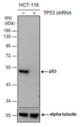
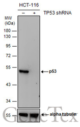
- Non-transfected (¡V) and transfected (+) HCT116 whole cell extracts (30 ?g) were separated by 10% SDS-PAGE, and the membrane was blotted with p53 antibody [D01] (GTX70214) diluted at 1:1000. The HRP-conjugated anti-mouse IgG antibody (GTX213111-01) was used to detect the primary antibody.
Supportive validation
- Submitted by
- GeneTex (provider)
- Main image
- Experimental details
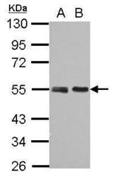
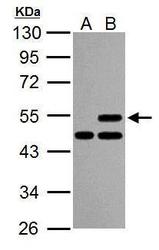
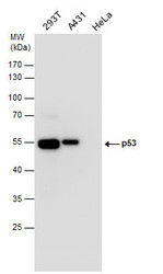
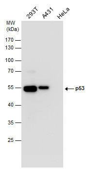
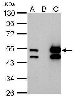
- Sample (30 ug of whole cell lysate) A: 293T B: A431 10% SDS PAGE GTX70214 diluted at 1:10000
- Validation comment
- WB

- Submitted by
- GeneTex (provider)
- Main image
- Experimental details
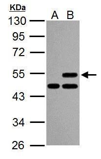
- Sample (30 ug of whole cell lysate) A: U2OS cells with mock treatment for 24hr B: U2OS cells with 30uM cisplatin treatment for 24hr 10% SDS PAGE GTX70214 diluted at 1:1000
- Validation comment
- WB
- Submitted by
- GeneTex (provider)
- Main image

- Experimental details
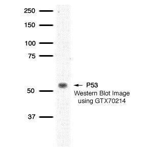
- Detection of human p53 protein using GeneTex p53 DO1 monoclonal antibody (GTX70214) in HBL100 whole cell extract.
- Validation comment
- WB
- Submitted by
- GeneTex (provider)
- Main image
- Experimental details
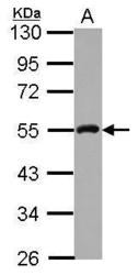
- Sample (30 ug of whole cell lysate) A: A431 10% SDS PAGE GTX70214 diluted at 1:5000
- Validation comment
- WB

- Submitted by
- GeneTex (provider)
- Main image
- Experimental details
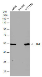
- p53 antibody detects p53 protein by western blot analysis. Various whole cell extracts (30 ?g) were separated by 10% SDS-PAGE, and the membrane was blotted with p53 antibody (GTX70214) diluted by 1:10000. The HRP-conjugated anti-mouse IgG antibody (GTX213111-01) was used to detect the primary antibody.
- Submitted by
- GeneTex (provider)
- Main image
- Experimental details
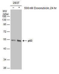
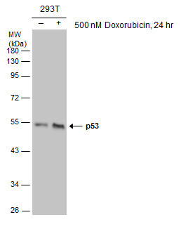
- Untreated (¡V) and treated (+) 293T whole cell extracts (30 ?g) were separated by 10% SDS-PAGE, and the membrane was blotted with p53 antibody [D01] (GTX70214) diluted at 1:1000. The HRP-conjugated anti-mouse IgG antibody (GTX213111-01) was used to detect the primary antibody.
- Submitted by
- GeneTex (provider)
- Main image
- Experimental details
- Non-transfected (¡V) and transfected (+) HCT116 whole cell extracts (30 ?g) were separated by 10% SDS-PAGE, and the membrane was blotted with p53 antibody [D01] (GTX70214) diluted at 1:1000. The HRP-conjugated anti-mouse IgG antibody (GTX213111-01) was used to detect the primary antibody.
- Submitted by
- GeneTex (provider)
- Main image
- Experimental details
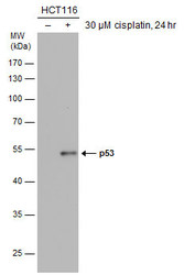
- Various whole cell extracts (30 ?g) were separated by 10% SDS-PAGE, and the membrane was blotted with p53 antibody [D01] (GTX70214) diluted at 1:1000. The HRP-conjugated anti-mouse IgG antibody (GTX213111-01) was used to detect the primary antibody.

- Submitted by
- GeneTex (provider)
- Main image
- Experimental details
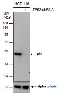
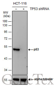
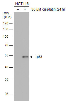
- Untreated (¡V) and treated (+) HCT116 whole cell extracts (30 ?g) were separated by 10% SDS-PAGE, and the membrane was blotted with p53 antibody [D01] (GTX70214) diluted at 1:1000. The HRP-conjugated anti-mouse IgG antibody (GTX213111-01) was used to detect the primary antibody.
Supportive validation
- Submitted by
- GeneTex (provider)
- Main image

- Experimental details
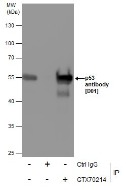
- Immunoprecipitation of p53 protein. HCT116 lysates with 30uM cisplatin treatment for 24 hours were subjected to immunoprecipitation using (B) normal mouse IgG or (C) 2.5 ug of anti-p53 antibody (GTX70214). (A) Input, 20ug of HCT116 lysate. The precipitated protein was detected by GTX70214 diluted at 1:10000. EasyBlot anti-Mouse IgG Kit (GTX225857-01) was used in Western blot.
- Validation comment
- IP
- Submitted by
- GeneTex (provider)
- Main image
- Experimental details
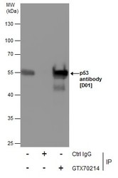
- Immunoprecipitation of p53 protein from 293T whole cell extracts using 5 £gg of p53 antibody [D01] (GTX70214).Western blot analysis was performed using p53 antibody [D01] (GTX70214).EasyBlot anti-Mouse IgG (GTX221667-01) was used as a secondary reagent.
Supportive validation
- Submitted by
- GeneTex (provider)
- Main image
- Experimental details
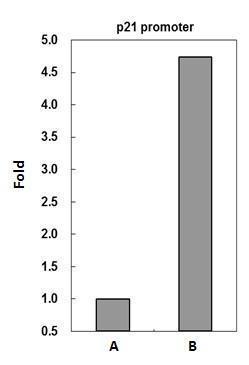
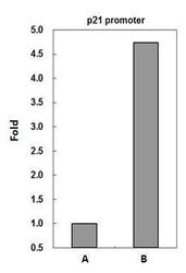
- p53 antibody immunoprecipitates p53 protein-DNA complex in ChIP experiments. ChIP Sample: HCT116 whole cell lysate/extract treated with CPT 500nM for 6hr A. 5 ?g preimmune mouse IgGB. 5 ?g of p53 antibody (GTX70214)The precipitated DNA was detected by PCR with primer set targeting to p21 promoter.
- Validation comment
- ChIP