 Explore
 Validate
 Learn
Learn Western blot
 Immunoprecipitation
Antibody data
- Antibody Data
- Antigen structure
- References [13]
- Comments [0]
- Validations
- Western blot [4]
- Immunohistochemistry [3]
- Flow cytometry [1]
- Other assay [5]
Submit
Validation data
Reference
Comment
Report error
- Product number
- MA5-12453 - Provider product page

- Provider
- Invitrogen Antibodies
- Product name
- p53 Monoclonal Antibody (PAb 122)
- Antibody type
- Monoclonal
- Antigen
- Other
- Description
- MA5-12453 targets p53 in FACS, IF, IP, and WB applications and shows reactivity with Hamster, Human, mouse, Non-human primate, and Rat samples. The MA5-12453 immunogen is sV40-transformed mouse B4 cells.
- Reactivity
- Human, Mouse, Rat, Hamster
- Host
- Mouse
- Isotype
- IgG
- Antibody clone number
- PAb 122
- Vial size
- 500 µL
- Concentration
- 0.2 mg/mL
- Storage
- 4° C
Submitted references Multi-Target Effects of the Cannabinoid CP55940 on Familial Alzheimer's Disease PSEN1 E280A Cholinergic-Like Neurons: Role of CB1 Receptor.
Telomerase activator-65 and pomegranate peel improved the health status of the liver in aged rats; multi-targets involved.
Cholinergic-like neurons carrying PSEN1 E280A mutation from familial Alzheimer's disease reveal intraneuronal sAPPβ fragments accumulation, hyperphosphorylation of TAU, oxidative stress, apoptosis and Ca2+ dysregulation: Therapeutic implications.
Establishment of three novel cell lines derived from African American patients with colorectal carcinoma: A unique tool for assessing racial health disparity.
PKM2 methylation by CARM1 activates aerobic glycolysis to promote tumorigenesis.
Simulated Microgravity Disrupts Cytoskeleton Organization and Increases Apoptosis of Rat Neural Crest Stem Cells Via Upregulating CXCR4 Expression and RhoA-ROCK1-p38 MAPK-p53 Signaling.
p53 acts as a co-repressor to regulate keratin 14 expression during epidermal cell differentiation.
A novel p53-binding domain in CUL7.
Targeting of p300/CREB binding protein coactivators by simian virus 40 is mediated through p53.
Gene expression and apoptosis induction in p53-heterozygous irradiated mice.
Iron deprivation induces apoptosis independently of p53 in human and murine tumour cells.
p53 associates with and targets Delta Np63 into a protein degradation pathway.
Retention of wild-type p53 in tumors from p53 heterozygous mice: reduction of p53 dosage can promote cancer formation.
Soto-Mercado V, Mendivil-Perez M, Jimenez-Del-Rio M, Velez-Pardo C
Journal of Alzheimer's disease : JAD 2021;82(s1):S359-S378
Journal of Alzheimer's disease : JAD 2021;82(s1):S359-S378
Telomerase activator-65 and pomegranate peel improved the health status of the liver in aged rats; multi-targets involved.
Alshinnawy AS, El-Sayed WM, Sayed AA, Salem AM, Taha AM
Iranian journal of basic medical sciences 2021 Jun;24(6):842-850
Iranian journal of basic medical sciences 2021 Jun;24(6):842-850
Cholinergic-like neurons carrying PSEN1 E280A mutation from familial Alzheimer's disease reveal intraneuronal sAPPβ fragments accumulation, hyperphosphorylation of TAU, oxidative stress, apoptosis and Ca2+ dysregulation: Therapeutic implications.
Soto-Mercado V, Mendivil-Perez M, Velez-Pardo C, Lopera F, Jimenez-Del-Rio M
PloS one 2020;15(5):e0221669
PloS one 2020;15(5):e0221669
Establishment of three novel cell lines derived from African American patients with colorectal carcinoma: A unique tool for assessing racial health disparity.
Paredes J, Ji P, Lacomb JF, Shroyer KR, Martello LA, Williams JL
International journal of oncology 2018 Oct;53(4):1516-1528
International journal of oncology 2018 Oct;53(4):1516-1528
PKM2 methylation by CARM1 activates aerobic glycolysis to promote tumorigenesis.
Liu F, Ma F, Wang Y, Hao L, Zeng H, Jia C, Wang Y, Liu P, Ong IM, Li B, Chen G, Jiang J, Gong S, Li L, Xu W
Nature cell biology 2017 Nov;19(11):1358-1370
Nature cell biology 2017 Nov;19(11):1358-1370
Simulated Microgravity Disrupts Cytoskeleton Organization and Increases Apoptosis of Rat Neural Crest Stem Cells Via Upregulating CXCR4 Expression and RhoA-ROCK1-p38 MAPK-p53 Signaling.
Lin SC, Gou GH, Hsia CW, Ho CW, Huang KL, Wu YF, Lee SY, Chen YH
Stem cells and development 2016 Aug 1;25(15):1172-93
Stem cells and development 2016 Aug 1;25(15):1172-93
p53 acts as a co-repressor to regulate keratin 14 expression during epidermal cell differentiation.
Cai BH, Hsu PC, Hsin IL, Chao CF, Lu MH, Lin HC, Chiou SH, Tao PL, Chen JY
PloS one 2012;7(7):e41742
PloS one 2012;7(7):e41742
A novel p53-binding domain in CUL7.
Kasper JS, Arai T, DeCaprio JA
Biochemical and biophysical research communications 2006 Sep 15;348(1):132-8
Biochemical and biophysical research communications 2006 Sep 15;348(1):132-8
Targeting of p300/CREB binding protein coactivators by simian virus 40 is mediated through p53.
Borger DR, DeCaprio JA
Journal of virology 2006 May;80(9):4292-303
Journal of virology 2006 May;80(9):4292-303
Gene expression and apoptosis induction in p53-heterozygous irradiated mice.
di Masi A, Antoccia A, Dimauro I, Argentino-Storino A, Mosiello A, Mango R, Novelli G, Tanzarella C
Mutation research 2006 Feb 22;594(1-2):49-62
Mutation research 2006 Feb 22;594(1-2):49-62
Iron deprivation induces apoptosis independently of p53 in human and murine tumour cells.
Truksa J, Kovár J, Valenta T, Ehrlichová M, Polák J, Naumann PW
Cell proliferation 2003 Aug;36(4):199-213
Cell proliferation 2003 Aug;36(4):199-213
p53 associates with and targets Delta Np63 into a protein degradation pathway.
Ratovitski EA, Patturajan M, Hibi K, Trink B, Yamaguchi K, Sidransky D
Proceedings of the National Academy of Sciences of the United States of America 2001 Feb 13;98(4):1817-22
Proceedings of the National Academy of Sciences of the United States of America 2001 Feb 13;98(4):1817-22
Retention of wild-type p53 in tumors from p53 heterozygous mice: reduction of p53 dosage can promote cancer formation.
Venkatachalam S, Shi YP, Jones SN, Vogel H, Bradley A, Pinkel D, Donehower LA
The EMBO journal 1998 Aug 17;17(16):4657-67
The EMBO journal 1998 Aug 17;17(16):4657-67
No comments: Submit comment
Supportive validation
- Submitted by
- Invitrogen Antibodies (provider)
- Main image

- Experimental details
- Western blot analysis was performed on whole cell extracts (30 µg lysate) of HEL 92.1.7 (Lane 1), MDA-MB-231 (Lane 2), COS-7 (Lane 3), A-431 (Lane 4), and SKOV3 (Lane 5). The blots were probed with Anti-p53 Mouse Monoclonal Antibody (Product # MA5-12453, 1-2 µg/mL) and detected by chemiluminescence using Goat anti-Mouse IgG (H+L) Secondary Antibody, HRP conjugate (Product # 62-6520, 1:4000 dilution). A 53 kDa band corresponding to p53 was observed across cell lines tested except SKOV3. An extra band at 100 kDa also observed. Known quantity of protein samples were electrophoresed using Novex® NuPAGE® 12 % Bis-Tris gel (Product # NP0342BOX), XCell SureLock™ Electrophoresis System (Product # EI0002) and Novex® Sharp Pre-Stained Protein Standard (Product # LC5800). Resolved proteins were then transferred onto a nitrocellulose membrane with iBlot® 2 Dry Blotting System (Product # IB21001). The membrane was probed with the relevant primary and secondary Antibody following blocking with 5 % skimmed milk. Chemiluminescent detection was performed using Pierce™ ECL Western Blotting Substrate (Product # 32106).

- Submitted by
- Invitrogen Antibodies (provider)
- Main image

- Experimental details
- Western blot analysis of p53 was performed by loading 20 µg of MCF-7 (lane 1) and MDA-MB-231 (lane 2) cell lysates in RIPA buffer (Product # 89901) and Page Ruler Plus Protein Ladder (Product # 26620) onto a 4-8% sodium dodecyl sulfate polyacrylamide gel (SDS-PAGE). Proteins were transferred to nitrocellulose membrane blocked in 5% milk /PBST for one hour at room temperature. p53 was detected using a cytochrome C monoclonal antibody (Product # MA5-12453) at a dilution of 1:500 5% milk /PBST overnight at at 4°C on a rocking platform, followed by a Goat anti-Mouse IgG (H + L) -HRP secondary antibody at a dilution of 1:5,000 for one hour. Chemiluminescent detection was performed using SuperSignal West Dura (Product # 34078) and Kodak M35 X-OMAT Automatic Processor. Data courtesy of Dr. Wei Xu at the University of Wisconsin, Madison, WI.

- Submitted by
- Invitrogen Antibodies (provider)
- Main image

- Experimental details
- Western blot was performed using Anti-p53 Monoclonal Antibody (PAb 122) (Product # MA5-12453) and a ~53 kDa band corresponding to TP53 was observed across cell lines tested . Whole cell extracts (30 µg lysate) of A-431 (Lane 1), T-47D (Lane 2), SK-O-V3 (Lane 3), HL-60 (Lane 4), U-937 (Lane 5) were electrophoresed using NuPAGE™ 4-12% Bis-Tris Protein Gel (Product # NP0321BOX). Resolved proteins were then transferred onto a nitrocellulose membrane (Product # IB23001) by iBlot® 2 Dry Blotting System (Product # IB21001). The blot was probed with the primary antibody (1:500 dilution) and detected by chemiluminescence with Goat anti-Mouse IgG (H+L) Superclonal™ Recombinant Secondary Antibody, HRP (Product # A28177,1:20000) using the iBright™ FL1500 Imaging System (Product # A44115). Chemiluminescent detection was performed using SuperSignal™ West Atto Ultimate Sensitivity Substrate (Product # A38556).

Supportive validation
- Submitted by
- Invitrogen Antibodies (provider)
- Main image

- Experimental details
- Immunohistochemistry analysis of p53 showing staining in the nucleus and weak staining in the cytoplasm of paraffin-embedded human breast carcinoma tissue (right) compared to a negative control without primary antibody (left). To expose target proteins, antigen retrieval was performed using 10mM sodium citrate (pH 6.0), microwaved for 8-15 min. Following antigen retrieval, tissues were blocked in 3% H2O2-methanol for 15 min at room temperature, washed with ddH2O and PBS, and then probed with a p53 Mouse Monoclonal Antibody (Product # MA5-12453) diluted in 3% BSA-PBS at a dilution of 1:20 for 1 hour at 37ºC in a humidified chamber. Tissues were washed extensively in PBST and detection was performed using an HRP-conjugated secondary antibody followed by colorimetric detection using a DAB kit. Tissues were counterstained with hematoxylin and dehydrated with ethanol and xylene to prep for mounting.

- Submitted by
- Invitrogen Antibodies (provider)
- Main image

- Experimental details
- Immunohistochemistry analysis of p53 showing staining in the nucleus and weak staining in the cytoplasm of paraffin-embedded human colon carcinoma tissue (right) compared to a negative control without primary antibody (left). To expose target proteins, antigen retrieval was performed using 10mM sodium citrate (pH 6.0), microwaved for 8-15 min. Following antigen retrieval, tissues were blocked in 3% H2O2-methanol for 15 min at room temperature, washed with ddH2O and PBS, and then probed with a p53 Mouse Monoclonal Antibody (Product # MA5-12453) diluted in 3% BSA-PBS at a dilution of 1:20 for 1 hour at 37ºC in a humidified chamber. Tissues were washed extensively in PBST and detection was performed using an HRP-conjugated secondary antibody followed by colorimetric detection using a DAB kit. Tissues were counterstained with hematoxylin and dehydrated with ethanol and xylene to prep for mounting.

- Submitted by
- Invitrogen Antibodies (provider)
- Main image

- Experimental details
- Immunohistochemistry analysis of p53 showing staining in the nucleus and weak staining in the cytoplasm of paraffin-embedded mouse colon tissue (right) compared to a negative control without primary antibody (left). To expose target proteins, antigen retrieval was performed using 10mM sodium citrate (pH 6.0), microwaved for 8-15 min. Following antigen retrieval, tissues were blocked in 3% H2O2-methanol for 15 min at room temperature, washed with ddH2O and PBS, and then probed with a p53 Mouse Monoclonal Antibody (Product # MA5-12453) diluted in 3% BSA-PBS at a dilution of 1:20 for 1 hour at 37ºC in a humidified chamber. Tissues were washed extensively in PBST and detection was performed using an HRP-conjugated secondary antibody followed by colorimetric detection using a DAB kit. Tissues were counterstained with hematoxylin and dehydrated with ethanol and xylene to prep for mounting.

Supportive validation
- Submitted by
- Invitrogen Antibodies (provider)
- Main image

- Experimental details
- Flow cytometry analysis of p53 was done on MDA-MB-231 cells. Cells were fixed with 70% ethanol for 10 minutes, permeabilized with 0.25% Triton™ X-100 for 20 minutes, and blocked with 5% BSA for 30 minutes at room temperature. Cells were labeled with p53 Mouse Monoclonal Antibody (MA512453, red histogram) or with mouse isotype control (yellow histogram) at 3-5 ug/million cells in 2.5% BSA. After incubation at room temperature for 2 hours, the cells were labeled with Alexa Fluor® 488 Rabbit Anti-Mouse Secondary Antibody (A11059) at a dilution of 1:400 for 30 minutes at room temperature. The representative 10,000 cells were acquired and analyzed for each sample using an Attune® Acoustic Focusing Cytometer. The purple histogram represents unstained control cells and the green histogram represents no-primary-antibody control.

Supportive validation
- Submitted by
- Invitrogen Antibodies (provider)
- Main image

- Experimental details
- Immunoprecipitation of p53 using p53 Monoclonal Antibody (Product # MA5-12453) on denatured Human MDA231 Cells.

- Submitted by
- Invitrogen Antibodies (provider)
- Main image
- Experimental details
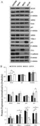
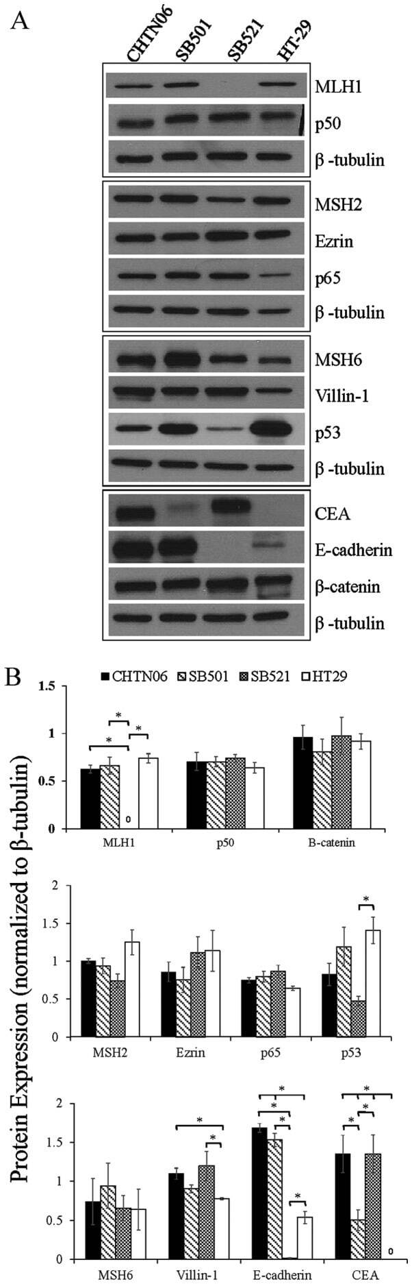
- Figure 3 The expression of proteins associated with colorectal carcinoma (CRC) tumorigenesis and metastasis was determined in the novel African American CRC lines by immunoblotting. (A) Qualitative analysis of CHTN06, SB501 and SB521 and HT-29, a Caucasian American CRC cell line, for protein expression of beta-catenin, p53, nuclear factor (NF)-kappaB (p50 and p65), villin-1, MSH2, MSH6, MLH1 and ezrin. (B) Semi-quantitative densitometry was performed by normalizing protein expression to the respective beta-tubulin loading control. Data were generated from three independent experiments. CEA, carcinoembryonic antigen.
- Submitted by
- Invitrogen Antibodies (provider)
- Main image

- Experimental details
- Fig 5 PSEN1 E280A ChLNs display activation of p53, PUMA, c-JUN and CASPASE-3. After 7 days of transdifferentiation, WT PSEN1 and PSEN1 E280A ChLNs were left in regular culture medium (RCm) for 0, 2 and 4 additional days, as indicated in the figure. After this time, the proteins in the extracts were blotted with primary antibodies against phosphorylated c-JUN (c-JUN), p53, PUMA, CASPASE-3 (CASP-3) and actin proteins. The intensities of the western blot bands shown in (A) were measured (B, C, D and E) by an infrared imaging system (Odyssey, LI-COR), and the intensity was normalized to that of actin. Additionally, after 4 days, ChLNs were double stained as indicated in the figure (F-I) with primary antibodies against p53 ( green ; F' and G') , PUMA ( red ; F"" and G"") , c-JUN ( green ; H' and I') and CASP-3 ( red ; H"" and I"") . The nuclei were stained with Hoechst 33342 ( blue ; F""'-I""') . (J-M) Quantification of p53 ( J ), PUMA ( K ), c-JUN ( L ) and CASP-3 ( M ) fluorescence intensity. Data are expressed as the mean +- SD; * p

- Submitted by
- Invitrogen Antibodies (provider)
- Main image

- Experimental details
- Fig. 4 CP55940 reduced the activation of p53, PUMA, c-Jun, and caspase-3 independent of CB 1 Rs signaling in PSEN1 E280A ChLNs. After 7 days of transdifferentiation, WT PSEN1 and PSEN1 E280A ChLNs were left Untreated or treated with SR, CP, or SR + CP in regular culture medium for 4 days. After this time, the proteins in the extracts were blotted with primary antibodies against phosphorylated c-Jun (p-c-JUN)/total c-Jun, p53, PUMA, caspase-3 (CASP-3) and actin proteins. The intensities of the western blot bands shown in (A) were measured (B-E) by an infrared imaging system (Odyssey, LI-COR), and the intensity was normalized to that of actin. Additionally, cells were double-stained as indicated in the figure (F-U) with primary antibodies against p53 ( green ; F'-M'), PUMA ( red ; F""-M""), c-JUN ( green ; N'-U'), and CASP-3 ( red ; N""-U""). The nuclei were stained with Hoechst 33342 ( blue ; F""'-U""'). V-X) Quantification of c-JUN (V), p53 (W), PUMA (X), and CASP-3 (Y) fluorescence intensity. Data are expressed as the mean+-SD; * p < 0.05; ** p < 0.01; *** p < 0.001. The blots and figures represent 1 out of 3 independent experiments. Image magnification, 200x.

- Submitted by
- Invitrogen Antibodies (provider)
- Main image

- Experimental details
- Figure 2 Effect of Ta-65 and pomegranate on the p53 protein (A-D, x200) and Caspase-3 activity (E) in liver tissue of rat (A) Control, (B) Aged, (C) Aged+Ta-65, and (D) Aged+pomegranate. Signals appear reddish-brown using DAB as a substrate, (E) Caspase-3 activity. Data are represented as Mean+-SD, n=8. Different letters denote significance at P< 0.05
