 Explore
 Validate
 Learn
Learn Western blot
 Immunoprecipitation
Antibody data
- Antibody Data
- Antigen structure
- References [4]
- Comments [0]
- Validations
- Western blot [1]
- Immunocytochemistry [1]
- Immunohistochemistry [2]
Submit
Validation data
Reference
Comment
Report error
- Product number
- AF1043 - Provider product page

- Provider
- R&D Systems
- Product name
- Human Phospho-p53 (S15) Antibody
- Antibody type
- Polyclonal
- Description
- Antigen Affinity-purified. Detects human, monkey, and rat p53 when phosphorylated at S15 and mouse p53 when phosphorylated at S18.
- Reactivity
- Human
- Host
- Rabbit
- Conjugate
- Unconjugated
- Isotype
- IgG
- Vial size
- 100 ug
- Concentration
- LYOPH
- Storage
- Use a manual defrost freezer and avoid repeated freeze-thaw cycles. 12 months from date of receipt, -20 to -70 °C as supplied. 1 month, 2 to 8 °C under sterile conditions after reconstitution. 6 months, -20 to -70 °C under sterile conditions after reconstitution.
Submitted references Paneth Cell Multipotency Induced by Notch Activation following Injury.
Skeletal muscle DNA damage precedes spinal motor neuron DNA damage in a mouse model of Spinal Muscular Atrophy (SMA).
Noninvasive assessment of regulable transferred-p53 gene expression and evaluation of therapeutic response with FDG-PET in tumor model.
The p53 oncoprotein is a substrate for tissue transglutaminase kinase activity.
Yu S, Tong K, Zhao Y, Balasubramanian I, Yap GS, Ferraris RP, Bonder EM, Verzi MP, Gao N
Cell stem cell 2018 Jul 5;23(1):46-59.e5
Cell stem cell 2018 Jul 5;23(1):46-59.e5
Skeletal muscle DNA damage precedes spinal motor neuron DNA damage in a mouse model of Spinal Muscular Atrophy (SMA).
Fayzullina S, Martin LJ
PloS one 2014;9(3):e93329
PloS one 2014;9(3):e93329
Noninvasive assessment of regulable transferred-p53 gene expression and evaluation of therapeutic response with FDG-PET in tumor model.
Aung W, Hasegawa S, Koshikawa-Yano M, Tsuji AB, Sogawa C, Sudo H, Sugyo A, Koizumi M, Furukawa T, Saga T
Gene therapy 2010 Sep;17(9):1142-51
Gene therapy 2010 Sep;17(9):1142-51
The p53 oncoprotein is a substrate for tissue transglutaminase kinase activity.
Mishra S, Murphy LJ
Biochemical and biophysical research communications 2006 Jan 13;339(2):726-30
Biochemical and biophysical research communications 2006 Jan 13;339(2):726-30
No comments: Submit comment
Supportive validation
- Submitted by
- R&D Systems (provider)
- Main image
- Experimental details
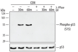
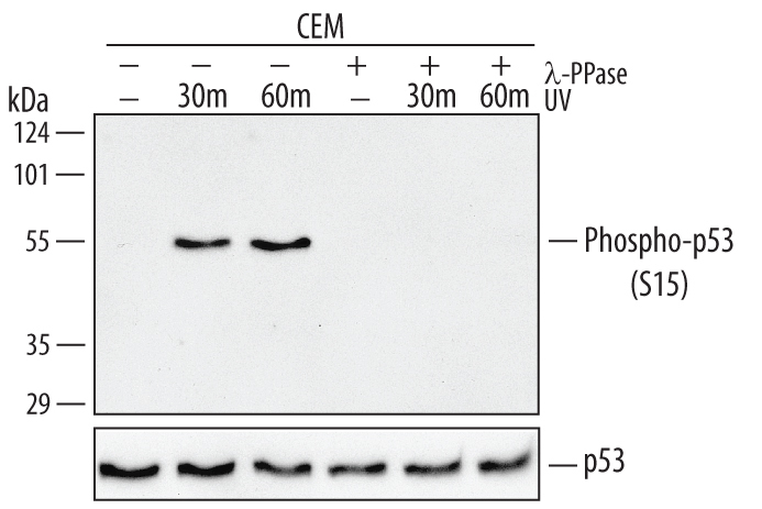
- Detection of Human Phospho-p53 (S15) by Western Blot. Western blot shows lysates of CEM human T-lymphoblastoid cell line untreated or exposed to 100 J/m2 UV-C for the indicated time. PVDF membrane was probed with 0.2 µg/mL Human Phospho-p53 (S15) Antigen Affinity-purified Polyclonal Antibody (Catalog # AF1043) followed by HRP-conjugated Anti-Rabbit IgG Secondary Antibody (Catalog # HAF008). A specific band for Phospho-p53 (S15) was detected at approximately 53 kDa (as indicated). The phospho-specificity of this antibody was supported by decreased labeling following treatment with 600 U lambda-phosphatase ( lambda-PPase) for 1 hour. For additional reference, duplicate lysates were probed with 1:5000 dilution Human/Mouse/Rat p53 HRP-conjugated Antigen Affinity-purified Polyclonal Antibody (lower panel, Catalog # HAF1355). This experiment was conducted under reducing conditions and using Immunoblot Buffer Group 1.
Supportive validation
- Submitted by
- R&D Systems (provider)
- Main image
- Experimental details
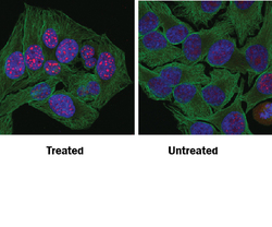
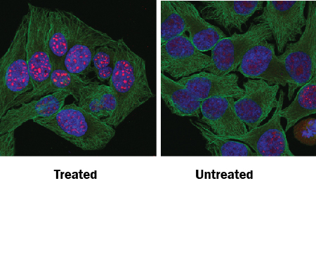
- Phospho-p53 (S15) in HeLa Human Cell Line. p53 phosphorylated at S15 was detected in immersion fixed HeLa human cervical epithelial carcinoma cell line treated with UV light (left panel) or untreated (right panel) using Rabbit Anti-Human Phospho-p53 (S15) Antigen Affinity-purified Polyclonal Antibody (Catalog # AF1043) at 10 µg/mL for 3 hours at room temperature. Cells were stained using the NorthernLights™ 557-conjugated Anti-Rabbit IgG Secondary Antibody (red; Catalog # NL004) and counterstained with DAPI (blue). Specific staining was localized to nuclei. Cells were co-stained using Rat Anti-Tubulin (Catalog # NB600-506, Novus Biologicals) and NorthernLights™ 493-conjugated Anti-Rat IgG Secondary Antibody (green; Catalog # NL015). View our protocol for Fluorescent ICC Staining of Cells on Coverslips.
Supportive validation
- Submitted by
- R&D Systems (provider)
- Main image
- Experimental details
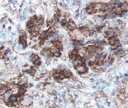
- Phospho-p53 (S15) in Human Breast Cancer Tissue. p53 phosphorylated at S15 was detected in immersion fixed paraffin-embedded sections of human breast cancer tissue using Human Phospho-p53 (S15) Antigen Affinity-purified Polyclonal Antibody (Catalog # AF1043) at 15 µg/mL overnight at 4 °C. Tissue was stained using the Anti-Rabbit HRP-DAB Cell & Tissue Staining Kit (brown; Catalog # CTS005) and counterstained with hematoxylin (blue). View our protocol for Chromogenic IHC Staining of immersion fixed paraffin-embedded Tissue Sections.

- Submitted by
- R&D Systems (provider)
- Main image
- Experimental details
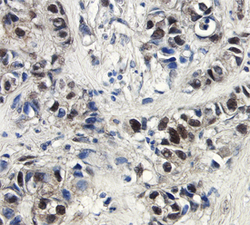
- Phospho-p53 (S15) in Human Breast Cancer Tissue. p53 phosphorylated at S15 was detected in immersion fixed paraffin-embedded sections of human breast cancer tissue using Human Phospho-p53 (S15) Antigen Affinity-purified Polyclonal Antibody (Catalog # AF1043) at 15 µg/mL overnight at 4 °C. Tissue was stained using the Anti-Rabbit HRP-DAB Cell & Tissue Staining Kit (brown; Catalog # CTS005) and counterstained with hematoxylin (blue). View our protocol for Chromogenic IHC Staining of immersion fixed paraffin-embedded Tissue Sections.
