Explore
 Validate
 Learn
Learn Immunohistochemistry
Antibody data
- Antibody Data
- Antigen structure
- References [2]
- Comments [0]
- Validations
- Immunohistochemistry [4]
Submit
Validation data
Reference
Comment
Report error
- Product number
- HPA028615 - Provider product page

- Provider
- Atlas Antibodies
- Proper citation
- Atlas Antibodies Cat#HPA028615, RRID:AB_10600717
- Product name
- Anti-INSL3
- Antibody type
- Polyclonal
- Reactivity
- Human
- Host
- Rabbit
- Conjugate
- Unconjugated
- Antigen sequence
EMREKLCGHHFVRALVRVCGGPRWSTEARRPATGG
DRELLQWLERRHLLHGLVADSNLTLGPGLQPLPQT
SHHHRHHRAAATNPARYCCLSGCTQQDLLTLCPY- Isotype
- IgG
- Vial size
- 100 µl
- Storage
- Store at +4°C for short term storage. Long time storage is recommended at -20°C.
Submitted references Cell context-specific expression of primary cilia in the human testis and ciliary coordination of Hedgehog signalling in mouse Leydig cells
The human testis-specific proteome defined by transcriptomics and antibody-based profiling
Nygaard M, Almstrup K, Lindbæk L, Christensen S, Svingen T
Scientific Reports 2015 May;5
Scientific Reports 2015 May;5
The human testis-specific proteome defined by transcriptomics and antibody-based profiling
Djureinovic D, Fagerberg L, Hallstrom B, Danielsson A, Lindskog C, Uhlen M, Ponten F
Molecular Human Reproduction 2014 May;20(6):476-488
Molecular Human Reproduction 2014 May;20(6):476-488
No comments: Submit comment
Enhanced validation
Supportive validation
- Submitted by
- Atlas Antibodies (provider)
- Enhanced method
- Orthogonal validation
- Main image
- Experimental details
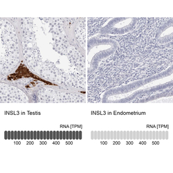
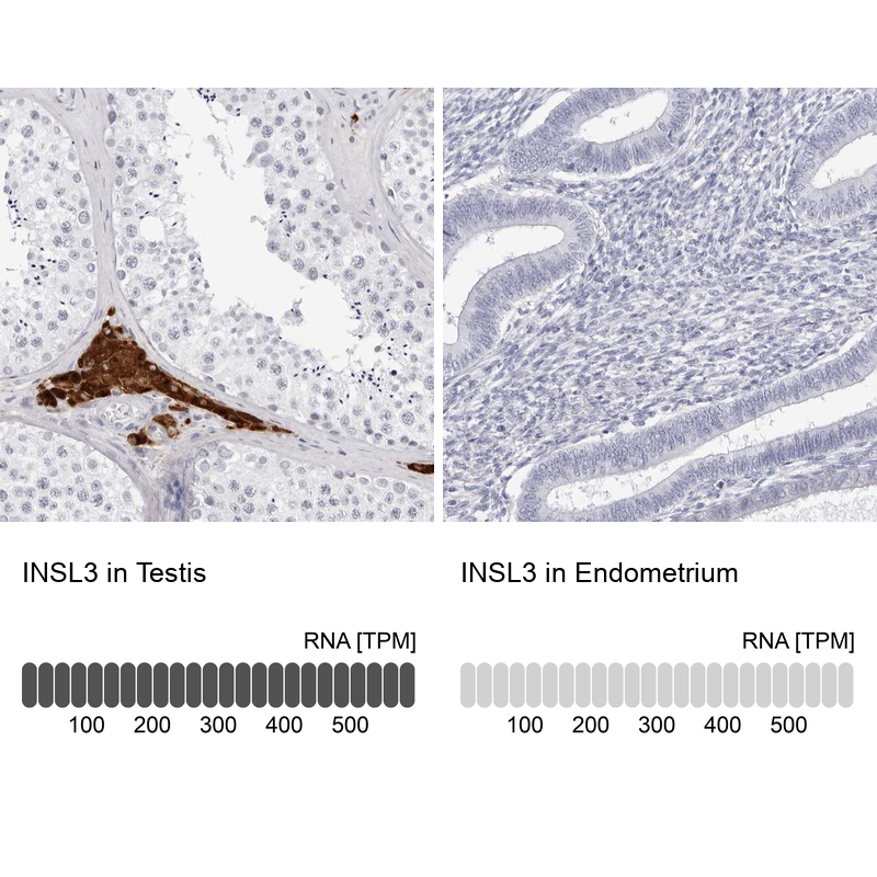
- Immunohistochemistry analysis in human testis and endometrium tissues using Anti-INSL3 antibody. Corresponding INSL3 RNA-seq data are presented for the same tissues.
- Sample type
- HUMAN
Supportive validation
- Submitted by
- Atlas Antibodies (provider)
- Main image
- Experimental details
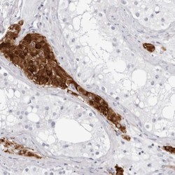
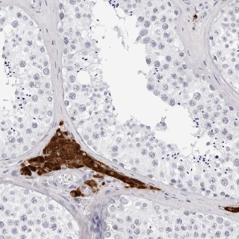
- Immunohistochemical staining of human testis shows strong cytoplasmic positivity in Leydig cells.

- Submitted by
- Atlas Antibodies (provider)
- Main image

- Experimental details
- Immunohistochemical staining of human testis shows high expression.
- Sample type
- HUMAN
- Submitted by
- Atlas Antibodies (provider)
- Main image

- Experimental details
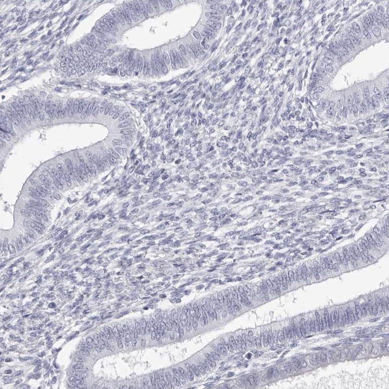
- Immunohistochemical staining of human endometrium shows low expression as expected.
- Sample type
- HUMAN