 Explore
 Validate
 Learn
Learn Western blot
Antibody data
- Antibody Data
- Antigen structure
- References [0]
- Comments [0]
- Validations
- Western blot [2]
- Flow cytometry [1]
Submit
Validation data
Reference
Comment
Report error
- Product number
- 700129 - Provider product page

- Provider
- Invitrogen Antibodies
- Product name
- Phospho-CREB (Ser133) Recombinant Rabbit Monoclonal Antibody (B16H16L26)
- Antibody type
- Monoclonal
- Antigen
- Other
- Reactivity
- Human, Mouse
- Host
- Rabbit
- Isotype
- IgG
- Antibody clone number
- B16H16L26
- Vial size
- 100 µg
- Concentration
- 0.5 mg/mL
- Storage
- Store at 4°C short term. For long term storage, store at -20°C, avoiding freeze/thaw cycles.
No comments: Submit comment
Supportive validation
- Submitted by
- Invitrogen Antibodies (provider)
- Main image

- Experimental details
- Western blot analysis of CREB (pS133) was performed by loading 20 µg of MDA-MB-231 (lane1), MCF7 (lane2), A549 (lane3), A431 (lane4) and HEK-293 (lane5) cell lysates using Novex®NuPAGE®4-12 % Bis-Tris gel (Product # NP0321BOX), XCell SureLock Electrophoresis System (Product # EI0002), Novex® Sharp Pre-Stained Protein Standard (Product # LC5800), and iBlot® Dry Blotting System (Product # IB21001). Proteins were transferred to a nitrocellulose membrane and blocked with 5 % skim milk for 1 hour at room temperature. CREB (ps133) was detected at ~43 kDa using CREB Recombinant Rabbit Monoclonal Antibody (Product # 700129) at 1-3 µg/mL in 2.5 % skim milk at 4°C overnight on a rocking platform. To confirm specificity, competition was performed with phosphopeptide (10 µg/mL) as shown in the corresponding blot on right. Goat anti-Rabbit IgG-HRP Secondary Antibody (Product # G-21234) at 1:5000 dilution was used and chemiluminescent detection was performed using Pierce™ ECL Western blotting Substrate (Product # 32106).

- Submitted by
- Invitrogen Antibodies (provider)
- Main image

- Experimental details
- Western blot analysis of CREB in NIH-3T3 cells treated with PDGF (30 µg) using a CREB recombinant rabbit monoclonal antibody (Product # 700129) at a dilution of 0.01 µg/mL. NBT/BCIP was used as the substrate (Product # WB7105). Results show a band at ~43 kDa. The second ~37 kDa band is seen in many cell types and is probably a close member of the ATF family. Competition assays show the disappearance of both the bands.

Supportive validation
- Submitted by
- Invitrogen Antibodies (provider)
- Main image
- Experimental details
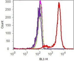
- Flow cytometry analysis of CREB [pS133] was done on HeLa cells. Cells were fixed with 70% ethanol for 10 minutes, permeabilized with 0.25% Tritonª X-100 for 20 minutes, and blocked with 5% BSA for 1 hour at room temperature. Cells were labeled with ABfinityª CREB [pS133] Recombinant Rabbit Monoclonal Antibody (700129, red histogram) or with rabbit isotype control (pink histogram) at 1-3 µg/million cells in 2.5% BSA. After incubation at room temperature for 2-3 hours, the cells were labeled with Alexa Fluor¨ 488 Goat Anti-Rabbit Secondary Antibody (A11008) at a dilution of 1:400 for 30 minutes at room temperature. The representative 10,000 cells were acquired and analyzed for each sample using an Attune¨ Acoustic Focusing Cytometer. The purple histogram represents unstained control cells and the green histogram represents no-primary-antibody control.
