 Explore
 Validate
 Learn
Learn Western blot
Antibody data
- Antibody Data
- Antigen structure
- References [2]
- Comments [0]
- Validations
- Western blot [4]
- Immunocytochemistry [1]
- Chromatin Immunoprecipitation [1]
- Other assay [2]
Submit
Validation data
Reference
Comment
Report error
- Product number
- 44-297G - Provider product page

- Provider
- Invitrogen Antibodies
- Product name
- Phospho-CREB (Ser129, Ser133) Polyclonal Antibody
- Antibody type
- Polyclonal
- Antigen
- Synthetic peptide
- Reactivity
- Human, Mouse
- Host
- Rabbit
- Isotype
- IgG
- Vial size
- 100 µL
- Storage
- -20°C
Submitted references Identification of a Novel Axon Regeneration Role for Noncanonical Wnt Signaling in the Adult Retina after Injury.
Rhynchosia volubilis Promotes Cell Survival via cAMP-PKA/ERK-CREB Pathway.
Musada GR, Carmy-Bennun T, Hackam AS
eNeuro 2022 Jul-Aug;9(4)
eNeuro 2022 Jul-Aug;9(4)
Rhynchosia volubilis Promotes Cell Survival via cAMP-PKA/ERK-CREB Pathway.
Ahn SH, Suh JS, Jang YK, Kim HS, Choi GH, Kim E, Kim TJ
Pharmaceuticals (Basel, Switzerland) 2022 Jan 6;15(1)
Pharmaceuticals (Basel, Switzerland) 2022 Jan 6;15(1)
No comments: Submit comment
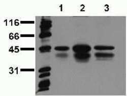
Supportive validation
- Submitted by
- Invitrogen Antibodies (provider)
- Main image

- Experimental details
- Whole cell extracts of human A549 cells (approximately 20,000 cells per lane) untreated (1), stimulated with EGF (2) or treated with pervanadate (3), were resolved by SDS-PAGE and transferred to PVDF. The membrane was blocked with a casein/Tween 20 buffer, then incubated with mouse anti-CREB/ATF1 (pS133) antibody (Product # 44-297G) at 0.5 µg/mL for 1 hour at room temperature. After washing, the membrane was incubated with an anti-mouse HRP-conjugated secondary antibody and signals were detected using an ECL detection method (exposure time: 30 seconds).

- Submitted by
- Invitrogen Antibodies (provider)
- Main image

- Experimental details
- Western blot analysis of CREB (pS129/pS133) was performed by loading 20 µg of NIH/3T3 (lane1), NIH/3T3 treated for 10 minutes with 50 ng/mL of PDGF (lane2), NIH/3T3 treated for 10 minutes with 200 ng/mL of EGF (lane3), A549 (lane4), A549 treated for 10 minutes with 200 ng/mL of EGF (lane5), A431 (lane6), SK-N-SH (lane7), MCF7 (lane8) and MDA-MB-231 (lane9) cell lysate using Novex®NuPAGE® 12 % Bis-Tris gel (Product # NP0342BOX), XCell SureLock Electrophoresis System (Product # EI0002), Novex® Sharp Pre-Stained Protein Standard (LC5800), and iBlot® 2 Dry Blotting System (IB21001). Proteins were transferred to a nitrocellulose membrane and blocked with 5% skim milk for 1 hour at room temperature. CREB (pS129/pS133) was detected at ~ 35 and 43 kDa using CREB (pS129/pS133) Rabbit Polyclonal Antibody (Product # 44-297G) at 1:1000 dilution in 5% skim milk at 4°C overnight on a rocking platform. Goat Anti-Rabbit IgG - HRP Secondary Antibody (G21234) at 1:5000 dilution was used and chemiluminescent detection was performed using Pierce™ ECL Western Blotting Substrate (Product # 32106).

- Submitted by
- Invitrogen Antibodies (provider)
- Main image

- Experimental details
- Peptide Competition and Phosphatase Treatment. Extracts of NIH3T3 cells untreated (1) or treated with 50 ng/mLPDGF for 15 minutes (2-6) were resolved by SDS-PAGE on a 10% Tris-glycine gel and transferred to PVDF. The membrane was blocked with a 5% BSA-TBST buffer for one hour at room temperature and either left untreated (1-5) or treated with lambda phosphatase (6), then incubated with the CREB [pSpS129/133] antibody (Product # 44-297G) for two hours at room temperature in a 3% BSA-TBST buffer, following prior incubation with: no peptide (1, 2, 6), the non-phosphopeptide corresponding to the phosphopeptide immunogen (3), a generic phosphoserine-containing peptide (4), or the phosphopeptide immunogen (5). After washing, the membrane was incubated with goat F(ab’)2 anti-rabbit IgG HRP conjugate (Product # ALI4404) and signals were detected using the Pierce SuperSignal™ method. The data show that only the phosphopeptide corresponding to CREB [pSpS129/133] blocks the antibody signal, demonstrating the specificity of the antibody. The data also show that phosphatase stripping eliminates the signal, further verifying that the antibody is phospho-specific. Note: Pretreatment of cells with PKA inhibitor (PKI) or GSK-3beta inhibitor (LiCl) diminished the signal, indicating the dual phosphoreactivity of this antibody (data not shown).

Supportive validation
- Submitted by
- Invitrogen Antibodies (provider)
- Main image

- Experimental details
- Immunofluorescent analysis of CREB (pS129/pS133) was done on 70% confluent log phase HeLa cells. The cells were fixed with 4% paraformaldehyde for 15 minutes, permeabilized with 0.25% Triton X-100 for 10 minutes, and blocked with 5% BSA for 1 hour at room temperature. The cells were labeled with CREB (pS129/pS133) Rabbit polyclonal Antibody (Product # 44-297G) at 2 µg/mL in 1% BSA and incubated for 3 hours at room temperature and then labeled with Alexa Fluor 488 Goat Anti-Rabbit IgG Secondary Antibody (Product # A-11008) at a dilution of 1:400 for 30 minutes at room temperature (Panel a: green). Nuclei (Panel b: blue) were stained with SlowFade® Gold Antifade Mountant with DAPI (Product # S36938). F-actin (Panel c: red) was stained with Alexa Fluor 594 Phalloidin (Product # A12381). Panel d is a merged image showing nuclear localization. Panel e shows no primary antibody control. The images were captured at 20X magnification.

Supportive validation
- Submitted by
- Invitrogen Antibodies (provider)
- Main image

- Experimental details
- ChIP- qPCR analysis of CREB (pSer133) was performed with 10 µL of the CREB (pSer133) Rabbit polyclonal antibody (Product # 44-297G) on sheared chromatin from 2 million HeLa cells treated with 50 ng/mL of TNFalpha for one hour using the MAGnify Chromatin Immunoprecipitation System (Product # 49-2024). Normal Rabbit IgG was used as a negative IP control. The purified DNA from each ChIP sample was analyzed by StepOnePlus Real-Time PCR System (Product # 4376600) with primers for the promoter of active YWHAZ, I-kB gene, used as positive control target, and the inactive GAPDH, used as negative control target. Data is presented as fold enrichment of the antibody signal versus the negative control IgG using the comparative CT method.

Supportive validation
- Submitted by
- Invitrogen Antibodies (provider)
- Main image

- Experimental details
- Figure 5 Effects of EERV on phosphorylated CREB activation. ( A ) RT-PCR assay shows mRNA expression levels of CREB in HeLa cells exposed to the control (0.5% ( v / v ) DMSO) and EERV (50 mug/mL) for 24 h. ( B ) The bar graphs represent mean values of relative CREB mRNA expression, and error bars indicate the S.E.M. (white and black; n = 3 each). ( C ) Phosphorylation of CREB was analyzed by Western blot with phospho-CREB antibodies and relative to total CREB protein after normalization to beta-tubulin levels. Protein expression levels of p-CREB, CREB, and beta-tubulin in HeLa cells exposed to control (0.5% ( v / v ) DMSO) and EERV (50 mug/mL) for 24 h. ( D - F ) The bar graph represents the mean values of relative p-CREB, CREB protein expression, and p-CREB/CREB expression ratios with error bars indicating the S.E.M. (white and black; n = 3 each, * p < 0.05, Student's t -test). Immunoreactive protein bands were detected using a 1:1 mixture of ProNA TM ECL Ottimo A and B (TransLab, Daejeon, Republic of Korea) and measured densitometrically using an iBright TM FL 1500 Imaging System (A44241, Invitrogen, Carlsbad, CA, USA).

- Submitted by
- Invitrogen Antibodies (provider)
- Main image

- Experimental details
- Figure 10. Increased phosphorylation CREB in Wnt5a-injected retinas. Upper panel shows representative images of cross-sections of saline-injected retinas. Lower panel shows representative images of cross-sections of 50 ng Wnt5a-injected retinas. IHC was performed to co-immunodetect phospho CREB (red) and RBPMS (green). Phosphorylation of CREB was induced in the GCL and other layers of the retina. White arrows indicate induced phosphorylation of CREB in RBPMS-positive RGC cell somas (GCL: ganglion cell layer, IPL: inner plexiform layer, INL: inner nuclear layer, OPL: outer plexiform layer, ONL: outer nuclear layer and OS: outer segment layer). Scale bar: 50 mum.
