 Explore
 Validate
 Learn
LearnHPA026761
antibody from Atlas Antibodies
Targeting: EPCAM
17-1A, 323/A3, CD326, CO-17A, EGP-2, EGP34, EGP40, Ep-CAM, ESA, GA733-2, HEA125, KS1/4, KSA, Ly74, M4S1, MH99, MIC18, MK-1, MOC31, TACST-1, TACSTD1, TROP1
 Western blot
Antibody data
- Antibody Data
- Antigen structure
- References [0]
- Comments [0]
- Validations
- Western blot [2]
- Immunohistochemistry [7]
Submit
Validation data
Reference
Comment
Report error
- Product number
- HPA026761 - Provider product page

- Provider
- Atlas Antibodies
- Proper citation
- Atlas Antibodies Cat#HPA026761, RRID:AB_1848198
- Product name
- Anti-EPCAM
- Antibody type
- Polyclonal
- Reactivity
- Human
- Host
- Rabbit
- Conjugate
- Unconjugated
- Antigen sequence
LFKAKQCNGTSMCWCVNTAGVRRTDKDTEITCSER
VRTYWIIIELKHKAREKPYDSKSLRTALQKEITTR
YQLDPKFITSILYENNVITIDLVQNSSQKTQNDVD
IADVAYY- Isotype
- IgG
- Vial size
- 100 µl
- Storage
- Store at +4°C for short term storage. Long time storage is recommended at -20°C.
No comments: Submit comment
Supportive validation
Enhanced validation
- Submitted by
- Atlas Antibodies (provider)
- Enhanced method
- Orthogonal validation
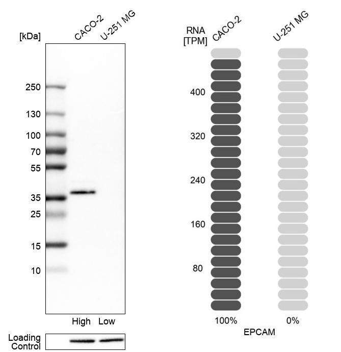
- Main image

- Experimental details
- Western blot analysis in human cell lines Caco-2 and U-251MG using Anti-EPCAM antibody. Corresponding EPCAM RNA-seq data are presented for the same cell lines. Loading control: Anti-PARP1.
Enhanced validation
- Submitted by
- Atlas Antibodies (provider)
- Enhanced method
- Recombinant expression validation
- Main image

- Experimental details
- Western blot analysis in control (vector only transfected HEK293T lysate) and EPCAM over-expression lysate (Co-expressed with a C-terminal myc-DDK tag (~3.1 kDa) in mammalian HEK293T cells, LY400847).
- Sample type
- HUMAN

Enhanced validation
Supportive validation
- Submitted by
- Atlas Antibodies (provider)
- Enhanced method
- Orthogonal validation
- Main image

- Experimental details
- Immunohistochemistry analysis in human small intestine and placenta tissues using HPA026761 antibody. Corresponding EPCAM RNA-seq data are presented for the same tissues.
- Sample type
- HUMAN

Supportive validation
- Submitted by
- Atlas Antibodies (provider)
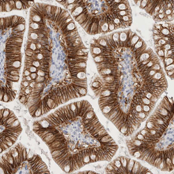
- Main image

- Experimental details
- Immunohistochemical staining of human rectum shows high expression.
- Sample type
- HUMAN

- Submitted by
- Atlas Antibodies (provider)
- Main image

- Experimental details
- Immunohistochemical staining of human liver shows low expression as expected.
- Sample type
- HUMAN

- Submitted by
- Atlas Antibodies (provider)
- Main image

- Experimental details
- Immunohistochemical staining of human Fallopian tube shows moderate membranous positivity in glandular cells.
- Sample type
- HUMAN

- Submitted by
- Atlas Antibodies (provider)
- Main image

- Experimental details
- Immunohistochemical staining of human kidney shows moderate membranous positivity in cells in tubules.
- Sample type
- HUMAN

- Submitted by
- Atlas Antibodies (provider)
- Main image

- Experimental details
- Immunohistochemical staining of human placenta shows no positivity in trophoblastic cells as expected.
- Sample type
- HUMAN

- Submitted by
- Atlas Antibodies (provider)
- Main image
- Experimental details
- Immunohistochemical staining of human small intestine shows strong membranous positivity in glandular cells.
- Sample type
- HUMAN
