Explore
 Validate
 Learn
Learn Western blot
Antibody data
- Antibody Data
- Antigen structure
- References [2]
- Comments [0]
- Validations
- Western blot [2]
- Immunocytochemistry [2]
- Flow cytometry [1]
Submit
Validation data
Reference
Comment
Report error
- Product number
- 44-454G - Provider product page

- Provider
- Invitrogen Antibodies
- Product name
- Phospho-MEK1/MEK2 (Ser218, Ser222, Ser226) Polyclonal Antibody
- Antibody type
- Polyclonal
- Antigen
- Synthetic peptide
- Reactivity
- Human, Mouse, Rat
- Host
- Rabbit
- Isotype
- IgG
- Vial size
- 100 µL
- Storage
- -20°C
Submitted references Fat1 deletion promotes hybrid EMT state, tumour stemness and metastasis.
Toll-like receptor-mediated production of IL-1Ra is negatively regulated by GSK3 via the MAPK ERK1/2.
Pastushenko I, Mauri F, Song Y, de Cock F, Meeusen B, Swedlund B, Impens F, Van Haver D, Opitz M, Thery M, Bareche Y, Lapouge G, Vermeersch M, Van Eycke YR, Balsat C, Decaestecker C, Sokolow Y, Hassid S, Perez-Bustillo A, Agreda-Moreno B, Rios-Buceta L, Jaen P, Redondo P, Sieira-Gil R, Millan-Cayetano JF, Sanmatrtin O, D'Haene N, Moers V, Rozzi M, Blondeau J, Lemaire S, Scozzaro S, Janssens V, De Troya M, Dubois C, Pérez-Morga D, Salmon I, Sotiriou C, Helmbacher F, Blanpain C
Nature 2021 Jan;589(7842):448-455
Nature 2021 Jan;589(7842):448-455
Toll-like receptor-mediated production of IL-1Ra is negatively regulated by GSK3 via the MAPK ERK1/2.
Rehani K, Wang H, Garcia CA, Kinane DF, Martin M
Journal of immunology (Baltimore, Md. : 1950) 2009 Jan 1;182(1):547-53
Journal of immunology (Baltimore, Md. : 1950) 2009 Jan 1;182(1):547-53
No comments: Submit comment
Supportive validation
- Submitted by
- Invitrogen Antibodies (provider)
- Main image

- Experimental details
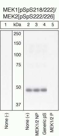
- Peptide Competition and Stimulation Extracts of HeLa cells untreated (1) or treated with 200 ng/mL PMA for 10 minutes (2-5) were resolved by SDS-PAGE on a 10% Tris-glycine gel and transferred to PVDF. The membrane was blocked with a 5% milk-TBST buffer for 1 hour at RT, then incubated with the MEK1 (pSpS218/222)/MEK2 (pSpS222/226) antibody at 4°C in a 3% milk-TBST buffer, following prior incubation with: no peptide (1, 2), the non-phosphopeptide corresponding to the phosphopeptide immunogen (3), a generic phosphoserine-containing peptide (4), or the phosphopeptide immunogen (5). After washing, the membrane was incubated with goat F (ab’)2 anti-rabbit IgG alkaline phosphatase (Product # ALI4405) and signals were detected using the Pierce SuperSignal™ method. The data show that only the phosphopeptide corresponding to MEK1 (pSpS218/222)/MEK2 (pSpS222/226) blocks the antibody signal, demonstrating the specificity of the antibody. The data also show the induction of MEK1 (pSpS218/222)/MEK2 (pSpS222/226) phosphorylation by the addition of PMA to this cell system.
- Submitted by
- Invitrogen Antibodies (provider)
- Main image
- Experimental details
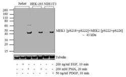
- Western blot analysis of MEK1 (pS218)/ (pS222) + MEK2 (pS222)/ (pS226) was performed by loading 20 µg of Jurkat (lane1), Jurkat treated for 10 minutes with 200 ng/mL of EGF (lane2), Jurkat treated for 20 minutes with 200 nM of PMA (lane3), HEK-293 (lane4), HEK-293 treated for 20 minutes with 200 nM of PMA (lane5), NIH\3T3 (lane6) and NIH\3T3 treated for 10 minutes with 50 ng/mL of PDGF (lane7) cell lysate using Novex® NuPAGE® 4-12 % Bis-Tris gel (Product # NP0322BOX), XCell SureLock™ Electrophoresis System (Product # EI0002), Novex® Sharp Pre-Stained Protein Standard (LC5800), and iBlot® 2 Dry Blotting System (IB21001). Proteins were transferred to a nitrocellulose membrane and blocked with 5 % skim milk for 1 hour at room temperature. MEK1 (pS218)/ (pS222) + MEK2 (pS222)/ (pS226) was detected at ~ 43 kDa using MEK1 (pS218)/ (pS222) + MEK2 (pS222)/ (pS226) Rabbit Polyclonal Antibody (Product # 44-244G) at 1:1000 dilution in 5 % skim milk at 4°C overnight on a rocking platform. Goat Anti-Rabbit IgG - HRP Secondary Antibody (G21234) at 1:5000 dilution was used and chemiluminescent detection was performed using Pierce™ ECL Western Blotting Substrate (Product # 32106).

Supportive validation
- Submitted by
- Invitrogen Antibodies (provider)
- Main image
- Experimental details
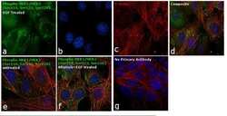
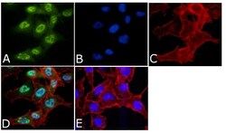
- Immunofluorescence analysis of Phospho- MEK1 (pSpS 218/222)/MEK2 (pSpS222/226) was performed using 70% confluent log phase NIH/3T3 cells treated with 200 ng/mL of EGF for 10 minutes. The cells were fixed with 4% paraformaldehyde for 10 minutes, permeabilized with 0.1% Triton™ X-100 for 10 minutes and blocked with 1% BSA for 1 hour at room temperature. The cells were labeled with Phospho- MEK1 (pSpS 218/222)/MEK2 (pSpS222/226) Rabbit Polyclonal Antibody (Product # 44-454G) at 5 µg/mL in 0.1% BSA and incubated overnight at 4 degree Celsius and then labeled with Goat anti-Rabbit IgG (H+L) Superclonal™ Secondary Antibody, Alexa Fluor® 488 conjugate (Product # A27034) at a dilution of 1:2000 for 45 minutes at room temperature (Panel a: green). Nuclei (Panel b: blue) were stained with SlowFade® Gold Antifade Mountant with DAPI (Product # S36938). F-actin (Panel c: red) was stained with Rhodamine Phalloidin (Product # R415, 1:300). Panel d represents the merged image showing cytoplasmic localization. Panel f represents cells treated with antagonist, Afatinib (1µM for 6hrs) followed by EGF (200 ng/mL for 10 minutes), showing reduced Phospho- MEK1 (pSpS 218/222)/MEK2 (pSpS222/226) staining. Panel e shows untreated cells with no signal. Panel g represents control cells with no primary antibody to assess background. The images were captured at 60X magnification.

- Submitted by
- Invitrogen Antibodies (provider)
- Main image
- Experimental details
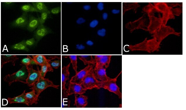
- Immunofluorescent analysis of Phospho- MEK1 (pSpS 218/222)/MEK2 (pSpS222/226) Antibody was done on 70% confluent log phase A549 cells. The cells were fixed with 4% paraformaldehyde for 15 minutes, permeabilized with 0.25% Triton™ X-100 for 10 minutes, and blocked with 5% BSA for 1 hour at room temperature. The cells were labeled with Phospho- MEK1 (pSpS 218/222)/MEK2 (pSpS222/226) Antibody (Product # 44-454G) at 1:250 dilution in 1% BSA and incubated for 3 hours at room temperature and then labeled with Alexa Fluor 488 Goat Anti-Rabbit IgG Secondary Antibody (Product # A-11008) at a dilution of 1:400 for 45 minutes at room temperature (Panel a: green). Nuclei (Panel b: blue) were stained with SlowFade® Gold Antifade Mountant with DAPI (Product # S36938). F-actin (Panel c: red) was stained with Alexa Fluor 594 Phalloidin (Product # A12381). Panel d is a merged image showing nuclear localization. Panel e is a no primary antibody control. The images were captured at 40X magnification.
Supportive validation
- Submitted by
- Invitrogen Antibodies (provider)
- Main image

- Experimental details
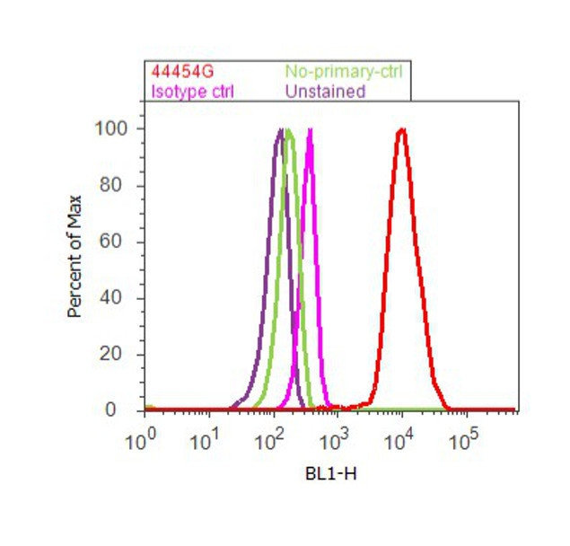
- Flow cytometry analysis of MEK1[pSpS 218/222]/MEK2 [pSpS222/226] was done on A549 cells treated with EGF (200ng/mL, 10 minutes). Cells were fixed with 70% ethanol for 10 minutes, permeabilized with 0.25% Triton™ X-100 for 20 minutes, and blocked with 5% BSA for 30 minutes at room temperature. Cells were labeled withMEK1[pSpS 218/222]/MEK2 [pSpS222/226] Rabbit Polyclonal Antibody (44454G, red histogram) or with rabbit isotype control (pink histogram) at 3-5 ug/million cells in 2.5% BSA. After incubation at room temperature for 2 hours, the cells were labeled with Alexa Fluor® 488 Goat Anti-Rabbit Secondary Antibody (A11008) at a dilution of 1:400 for 30 minutes at room temperature. The representative 10,000 cells were acquired and analyzed for each sample using an Attune® Acoustic Focusing Cytometer. The purple histogram represents unstained control cells and the green histogram represents no-primary-antibody control.