 Explore
 Validate
 Learn
LearnPA5-34803
antibody from Invitrogen Antibodies
Targeting: PARP1
ADPRT, ARTD1, PARP, PPOL
 Western blot Immunocytochemistry
 Immunoprecipitation Immunohistochemistry Chromatin Immunoprecipitation Other assay
Antibody data
- Antibody Data
- Antigen structure
- References [1]
- Comments [0]
- Validations
- Western blot [7]
- Immunocytochemistry [3]
- Immunohistochemistry [3]
- Chromatin Immunoprecipitation [3]
- Other assay [2]
Submit
Validation data
Reference
Comment
Report error
- Product number
- PA5-34803 - Provider product page

- Provider
- Invitrogen Antibodies
- Product name
- PARP1 Polyclonal Antibody
- Antibody type
- Polyclonal
- Antigen
- Recombinant protein fragment
- Description
- Recommended positive controls: 293T, A431, HeLa, HepG2, HCT116, HCT116 (30 µM cisplatin treatment for 24 hr), NIH-3T3, PC-12, H1299. Predicted reactivity: Mouse (90%), Rat (88%), Bovine (91%). Store product as a concentrated solution. Centrifuge briefly prior to opening the vial.
- Reactivity
- Human, Mouse, Rat
- Host
- Rabbit
- Isotype
- IgG
- Vial size
- 100 µL
- Concentration
- 0.48 mg/mL
- Storage
- Store at 4°C short term. For long term storage, store at -20°C, avoiding freeze/thaw cycles.
Submitted references Expanding the editable genome and CRISPR-Cas9 versatility using DNA cutting-free gene targeting based on in trans paired nicking.
Chen X, Tasca F, Wang Q, Liu J, Janssen JM, Brescia MD, Bellin M, Szuhai K, Kenrick J, Frock RL, Gonçalves MAFV
Nucleic acids research 2020 Jan 24;48(2):974-995
Nucleic acids research 2020 Jan 24;48(2):974-995
No comments: Submit comment
Supportive validation
- Submitted by
- Invitrogen Antibodies (provider)
- Main image

- Experimental details
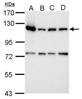
- Western blot analysis of PARP1 using 30 µg of A) 293T (B) A431 (C) HeLa and D) HepG2 lysate. Samples were loaded onto a 7.5% SDS-PAGE gel and probed with a PARP1 polyclonal antibody (Product # PA5-34803) at a dilution of 1:10,000.
- Submitted by
- Invitrogen Antibodies (provider)
- Main image

- Experimental details
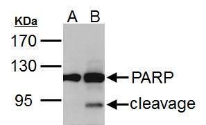
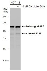
- Western blot analysis of PARP1 using A) 30 µg HCT116 whole cell lysate (untreated) and B) 30 µg HCT116 whole cell lysate (30uM cisplatin treatment for 24hr). Samples were loaded onto a 7.5% SDS-PAGE gel and probed with a PARP1 polyclonal antibody (Product # PA5-34803) at a dilution of 1:5000.
- Submitted by
- Invitrogen Antibodies (provider)
- Main image

- Experimental details
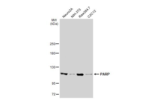
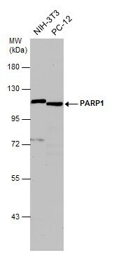
- Western Blot analysis of PARP1 was performed by separating 30 µg of various whole cell extracts by 5% SDS-PAGE. Proteins were transferred to a membrane and probed with a PARP1 Polyclonal Antibody (Product # PA5-34803) at a dilution of 1:1000 and a HRP-conjugated anti-rabbit IgG secondary antibody.

- Submitted by
- Invitrogen Antibodies (provider)
- Main image

- Experimental details
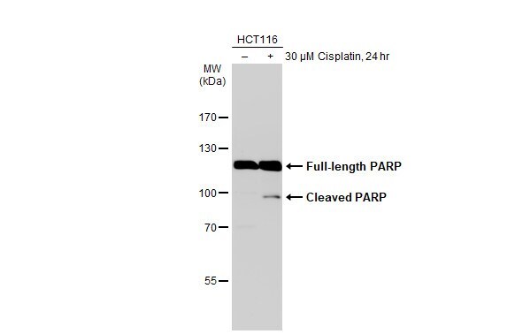
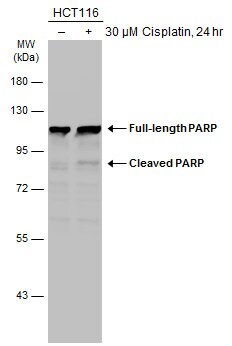
- Western Blot using PARP1 Polyclonal Antibody (Product # PA5-34803). Untreated (–) and treated (+) HCT116 whole cell extracts (30 µg) were separated by 7.5% SDS-PAGE, and the membrane was blotted with PARP1 Polyclonal Antibody (Product # PA5-34803) diluted at 1:1,000. The HRP-conjugated anti-rabbit IgG antibody was used to detect the primary antibody.
- Submitted by
- Invitrogen Antibodies (provider)
- Main image

- Experimental details
- Western Blot analysis of PARP1 was performed by separating 30 µg of various whole cell extracts by 5% SDS-PAGE. Proteins were transferred to a membrane and probed with a PARP1 Polyclonal Antibody (Product # PA5-34803) at a dilution of 1:1000 and a HRP-conjugated anti-rabbit IgG secondary antibody.
- Submitted by
- Invitrogen Antibodies (provider)
- Main image
- Experimental details
- Western Blot analysis of PARP1 was performed by separating 30 µg of untreated (–) and treated (+) HCT116 whole cell extracts by 7.5% SDS-PAGE. Proteins were transferred to a membrane and probed with a PARP1 Polyclonal Antibody (Product # PA5-34803) at a dilution of 1:1000.
- Submitted by
- Invitrogen Antibodies (provider)
- Main image

- Experimental details
- Western Blot analysis of PARP1 was performed by separating 30 µg of various whole cell extracts by 7.5% SDS-PAGE. Proteins were transferred to a membrane and probed with a PARP1 Polyclonal Antibody (Product # PA5-34803) at a dilution of 1:500 and a HRP-conjugated anti-rabbit IgG secondary antibody.
Supportive validation
- Submitted by
- Invitrogen Antibodies (provider)
- Main image
- Experimental details
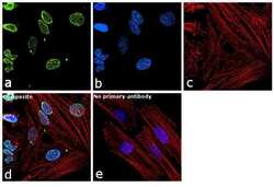
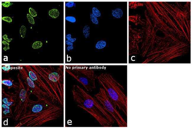
- Immunofluorescence analysis of PARP was performed using log phase HeLa cells. The cells were fixed with 4% paraformaldehyde for 10 minutes, permeabilized with 0.1% Triton™ X-100 for 10 minutes, and blocked with 1% BSA for 1 hour at room temperature. The cells were labeled with PARP Polyclonal Antibody (Product # PA5-34803) at 5 µg/mL in 0.1% BSA and incubated overnight at 4 degree and then labeled with Goat anti-Rabbit IgG (H+L) Superclonal™ Secondary Antibody, Alexa Fluor® 488 conjugate (Product # A27034) at a dilution of 1:2000 for 45 minutes at room temperature (Panel a: green). Nuclei (Panel b: blue) were stained with SlowFade® Gold Antifade Mountant with DAPI (Product # S36938). F-actin (Panel c: red) was stained with Rhodamine Phalloidin (Product # R415, 1:300). Panel d represents the merged image showing nuclear localization of PARP. Panel e represents control cells with no primary antibody to assess background. The images were captured at 60X magnification.
- Submitted by
- Invitrogen Antibodies (provider)
- Main image
- Experimental details
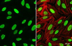
- Immunocytochemistry-Immunofluorescence analysis of PARP1 was performed in HeLa cells fixed in 4% paraformaldehyde at RT for 15 min. Green: PARP1 Polyclonal Antibody (Product # PA5-34803) diluted at 1:500. Red: phalloidin, a cytoskeleton marker. Scale bar = 10 µm.
- Submitted by
- Invitrogen Antibodies (provider)
- Main image
- Experimental details
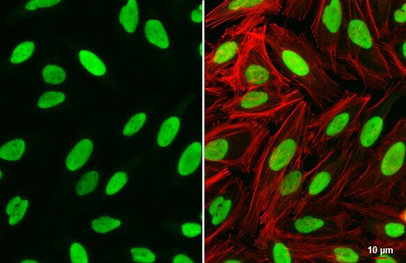
- PARP1 Polyclonal Antibody detects PARP protein at nucleus by immunofluorescent analysis. Sample: HeLa cells were fixed in 4% paraformaldehyde at RT for 15 min. Green: PARP stained by PARP1 Polyclonal Antibody (Product # PA5-34803) diluted at 1:500. Red: phalloidin, a cytoskeleton marker, diluted at 1:200. Scale bar= 10 µm.

Supportive validation
- Submitted by
- Invitrogen Antibodies (provider)
- Main image

- Experimental details
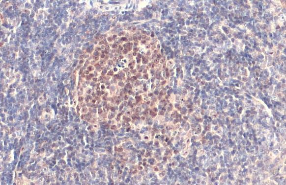
- PARP1 Polyclonal Antibody detects PARP protein at nucleus by immunohistochemical analysis. Sample: Paraffin-embedded human lung cancer. PARP stained by PARP1 Polyclonal Antibody (Product # PA5-34803) diluted at 1:500. Antigen Retrieval: Citrate buffer, pH 6.0, 15 min.

- Submitted by
- Invitrogen Antibodies (provider)
- Main image

- Experimental details
- PARP1 Polyclonal Antibody detects PARP protein at nucleus by immunohistochemical analysis. Sample: Paraffin-embedded mouse spleen. PARP stained by PARP1 Polyclonal Antibody (Product # PA5-34803) diluted at 1:500. Antigen Retrieval: Citrate buffer, pH 6.0, 15 min.
- Submitted by
- Invitrogen Antibodies (provider)
- Main image
- Experimental details
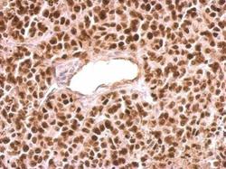
- PARP1 antibody [N2C1], Internal detects PARP1 protein at nucleus on HeLa xenograft by immunohistochemical analysis. Sample: Paraffin-embedded HeLa xenograft. PARP1 antibody [N2C1], Internal (Product # PA5-34803) dilution: 1:500. Antigen Retrieval: EDTA based buffer, pH 8.0, 15 min.

Supportive validation
- Submitted by
- Invitrogen Antibodies (provider)
- Main image

- Experimental details
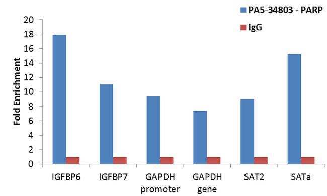
- Enrichment of endogenous PARP protein at specific gene loci using Anti-PARP Antibody: Chromatin Immunoprecipitation (ChIP) was performed using Anti-PARP Rabbit Polyclonal Antibody (Product # PA5-34803, 4 ug) on sheared chromatin from 2 million HeLa cells treated with Etoposide (50 uM for 6 hours) using the MAGnify ChIP System (Product # 49-2024). Normal Rabbit IgG was used as a negative IP control. The purified DNA was analyzed by qPCR with PCR primer pairs over IGFBP6, IGFBP7, GAPDH promoter and gene, SAT2 satellite repeats and SAT alpha. Data is presented as fold enrichment of the antibody signal versus the negative control IgG using the comparative CT method.
- Submitted by
- Invitrogen Antibodies (provider)
- Main image
- Experimental details
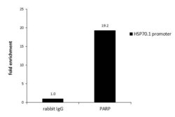
- ChIP assay analysis of PARP1 was performed in HeLa chromatin extract using 5 µg of either normal rabbit IgG or PARP1 Polyclonal Antibody (Product # PA5-34803). The precipitated DNA was detected by PCR with primer set targeting to HSP70.1 promoter.

- Submitted by
- Invitrogen Antibodies (provider)
- Main image

- Experimental details
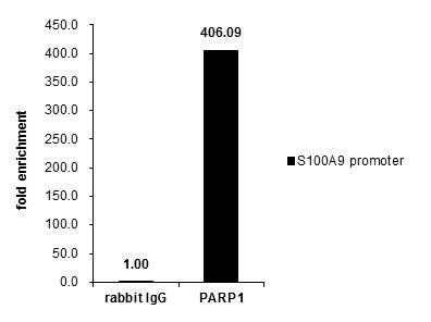
- Cross-linked ChIP was performed with Raji chromatin extract and 5 µg of either control rabbit IgG or PARP1 Polyclonal Antibody (Product # PA5-34803). The precipitated DNA was detected by PCR with primer set targeting to S100A9 promoter.
Supportive validation
- Submitted by
- Invitrogen Antibodies (provider)
- Main image
- Experimental details
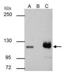
- PARP1 antibody [N2C1], Internal immunoprecipitates PARP1 protein in IP experiments. IP samples: HCT-116 whole cell extract. A. 30 µg HCT-116 whole cell extract. B. Control with 4 µg of preimmune Rabbit IgG. C. Immunoprecipitation of PARP1 protein by 4 µg PARP1 antibody [N2C1], Internal (Product # PA5-34803). 5 % SDS-PAGE. The immunoprecipitated PARP1 protein was detected by PARP1 antibody [N2C1], Internal (Product # PA5-34803) diluted at 1:3000.

- Submitted by
- Invitrogen Antibodies (provider)
- Main image
- Experimental details
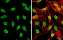
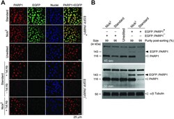
- Figure 5. Examination of PARP1 protein status after gene editing triggered by DSBs versus SSBs. ( A ) Confocal microscopy analysis of HeLa cells expressing untagged and EGFP-tagged PARP1. Confocal microscopy of EGFP::PARP1 + and EGFP::PARP1 - cells confirming co-localization of PARP1 and EGFP in the nuclei of the former cell populations engineered by in trans paired nicking or standard gene editing. Nuclei were counterstained with DAPI. Unedited HeLa cells served as negative controls. Specimens of EGFP::PARP1 - cells not incubated with the primary PARP1-specific antibody (-1st Ab) provided for an additional staining control. ( B ) Western blot analysis of HeLa cells expressing untagged and EGFP-tagged PARP1. Western blotting of EGFP::PARP1 + and EGFP::PARP1 - cells exposing a striking reduction in the amounts of endogenous PARP1 antigen exclusively in EGFP::PARP1 + cells generated through standard DSB-dependent gene editing (open arrowhead). Properly sized EGFP::PARP1 fusion products were detected in both EGFP::PARP1 + cell populations (solid arrowhead). Unedited HeLa cells served as negative controls. alpha/beta Tubulin antigens served as internal protein loading controls.
