Explore
 Validate
 Learn
Learn Western blot
 Immunoprecipitation
Antibody data
- Antibody Data
- Antigen structure
- References [0]
- Comments [0]
- Validations
- Western blot [4]
- Immunocytochemistry [1]
- Immunohistochemistry [1]
- Other assay [1]
Submit
Validation data
Reference
Comment
Report error
- Product number
- PA5-85676 - Provider product page

- Provider
- Invitrogen Antibodies
- Product name
- PKR Polyclonal Antibody
- Antibody type
- Polyclonal
- Antigen
- Recombinant full-length protein
- Reactivity
- Human
- Host
- Rabbit
- Isotype
- IgG
- Vial size
- 100 µL
- Concentration
- 1 mg/mL
- Storage
- Store at 4°C short term. For long term storage, store at -20°C, avoiding freeze/thaw cycles.
No comments: Submit comment
Supportive validation
- Submitted by
- Invitrogen Antibodies (provider)
- Main image
- Experimental details
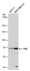
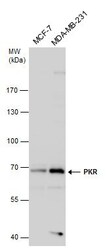
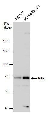
- Western blot analysis of PKR in various whole cell extracts using PKR polyclonal antibody (Product # PA5-85676) using 30 µg of sample at a dilution of 1:1000. Prior to incubation with primary antibody, the sample was separated on 7.5% SDS-PAGE.

- Submitted by
- Invitrogen Antibodies (provider)
- Main image
- Experimental details
- PKR Polyclonal Antibody detects PKR protein by western blot analysis. Various whole cell extracts (30 µg) were separated by 7.5% SDS-PAGE, and the membrane was blotted with PKR Polyclonal Antibody (Product # PA5-85676) diluted at a dilution of 1:1,000.
- Submitted by
- Invitrogen Antibodies (provider)
- Main image
- Experimental details
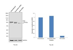
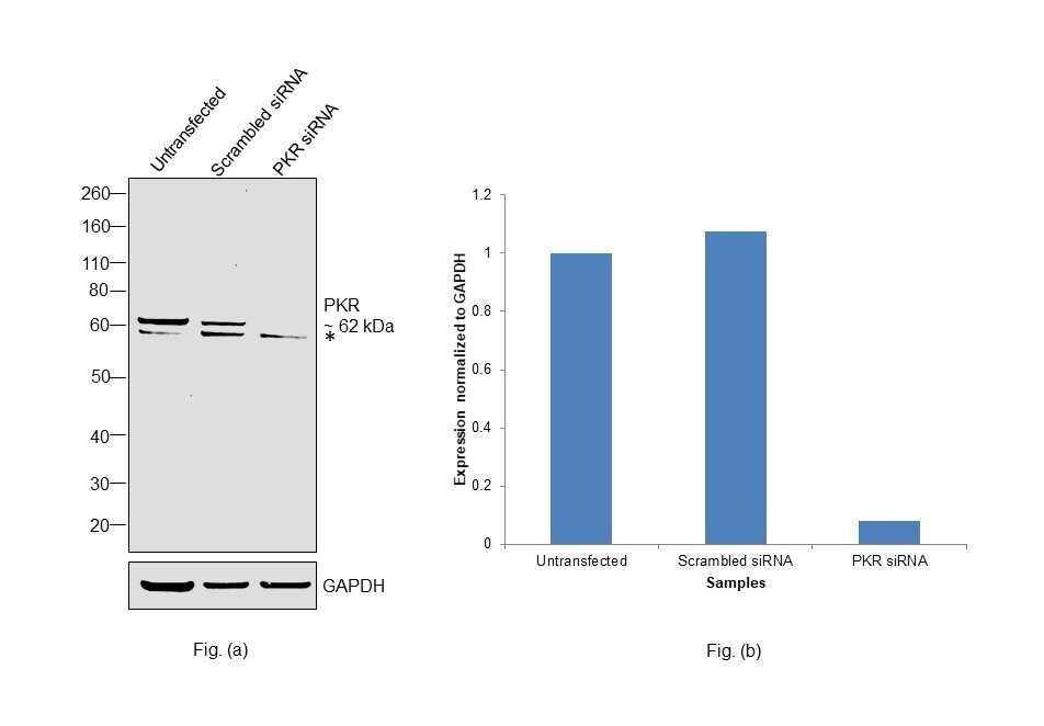
- Knockdown of Interferon-induced, double-stranded RNA-activated protein kinase was achieved by transfecting MCF7 with Interferon-induced, double-stranded RNA-activated protein kinase specific siRNAs (Silencer® select Product # S11187, S11185). Western Blot analysis (Fig. a) was performed using Whole cell extracts from the Interferon-induced, double-stranded RNA-activated protein kinase knockdown cells (lane 3), non-targeting scrambled siRNA transfected cells (lane 2) and untransfected cells (lane 1). The blot was probed with PKR Polyclonal Antibody (Product # PA5-85676, 1:1000 dilution) and Goat anti-Rabbit IgG (H+L) Superclonal™ Recombinant Secondary Antibody, HRP (Product # A27036, 1:20000 dilution). Densitometric analysis of this Western Blot is shown in histogram (Fig. b). Decrease in signal upon siRNA mediated knock down confirms that antibody is specific to Interferon-induced, double-stranded RNA-activated protein kinase.
- Submitted by
- Invitrogen Antibodies (provider)
- Main image
- Experimental details
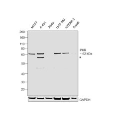
- Western Blot was performed using Anti-PKR Polyclonal Antibody (Product # PA5-85676) and a 62 kDa band corresponding to Interferon-induced, double-stranded RNA-activated protein kinase was observed across samples along with an uncharacterised (*) band at ~59 kDa. Whole cell extracts (40 µg lysate) of MCF7 (Lane 1), A-431 (Lane 2), A549 (Lane 3), U-87 MG (Lane 4), NTERA-2 cl.D1 (Lane 5), Daudi (Lane 6) were electrophoresed using NuPAGE™ 4-12% Bis-Tris Protein Gel (Product # NP0321BOX). Resolved proteins were then transferred onto a nitrocellulose membrane (Product # IB23001) by iBlot® 2 Dry Blotting System (Product # IB21001). The blot was probed with the primary antibody (1:1000 dilution) and detected by chemiluminescence with Goat anti-Rabbit IgG (H+L) Superclonal™ Recombinant Secondary Antibody, HRP (Product # A27036, 1:20000 dilution) using the iBright FL 1000 (Product # A32752). Chemiluminescent detection was performed using SuperSignal™ West Dura Extended Duration Substrate (Product # 34076).

Supportive validation
- Submitted by
- Invitrogen Antibodies (provider)
- Main image

- Experimental details
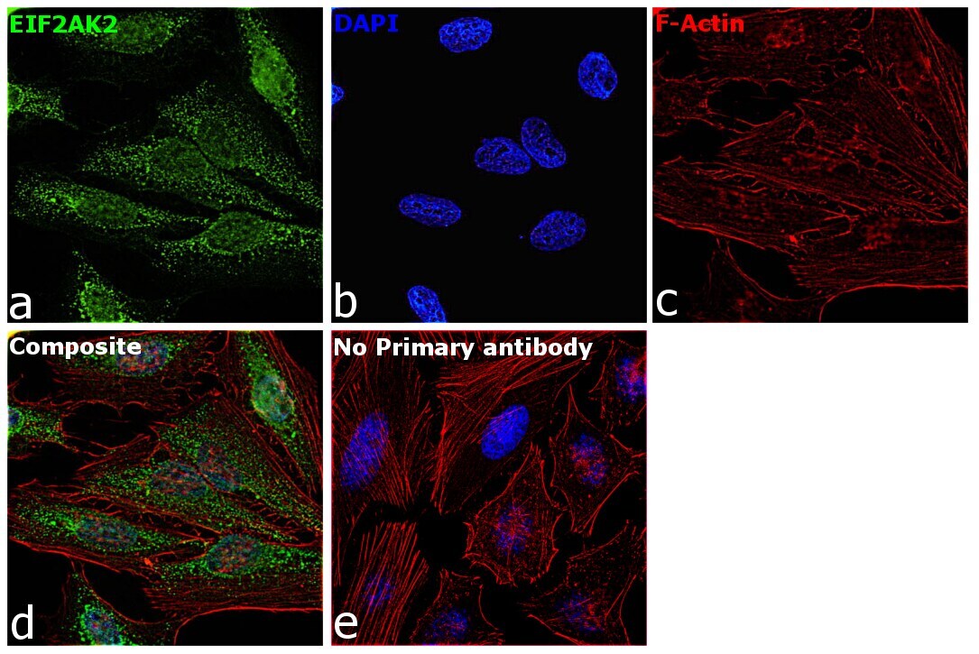
- Immunofluorescence analysis of Interferon-induced, double-stranded RNA-activated protein kinase was performed using 70% confluent log phase HeLa cells. The cells were fixed with 4% paraformaldehyde for 5 minutes, permeabilized with 0.1% Triton™ X-100 for 10 minutes, and blocked with 2% BSA for overnight at room temperature. The cells were labeled with PKR Polyclonal Antibody (Product # PA5-85676) at 1:100 dilution in 0.1% BSA, incubated at 4 degree celsius overnight and then labeled with Donkey anti-Rabbit IgG (H+L) Highly Cross-Adsorbed Secondary Antibody, Alexa Fluor Plus 488 (Product # A32790), (1:2000 dilution), for 45 minutes at room temperature (Panel a: Green). Nuclei (Panel b: Blue) were stained with ProLong™ Diamond Antifade Mountant with DAPI (Product # P36962). F-actin (Panel c: Red) was stained with Rhodamine Phalloidin (Product # R415, 1:300). Panel d represents the merged image showing cytoplasm and nucleus localization. Panel e represents control cells with no primary antibody to assess background. The images were captured at 60X magnification.
Supportive validation
- Submitted by
- Invitrogen Antibodies (provider)
- Main image
- Experimental details
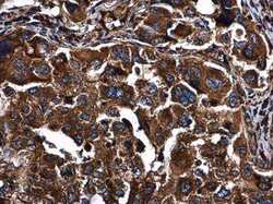
- PKR Polyclonal Antibody detects PKR protein at cytoplasm in human lung cancer by immunohistochemical analysis. Sample: Paraffin-embedded human lung cancer. PKR Polyclonal Antibody (Product # PA5-85676) diluted at 1:500. Antigen Retrieval: Citrate buffer, pH 6.0, 15 min.

Supportive validation
- Submitted by
- Invitrogen Antibodies (provider)
- Main image

- Experimental details
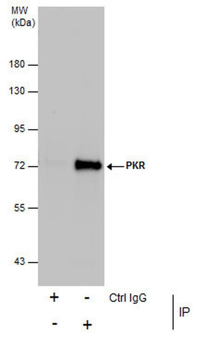
- Immunoprecipitation analysis of PKR in MCF-7 whole cell extracts with PKR polyclonal antibody (Product # PA5-85676) using 5 µg of sample, followed by anti-Rabbit IgG secondary antibody.