Explore
 Validate
 Learn
Learn Western blot
Antibody data
- Antibody Data
- Antigen structure
- References [0]
- Comments [0]
- Validations
- Western blot [1]
- Immunocytochemistry [1]
- Immunohistochemistry [5]
- Other assay [1]
Submit
Validation data
Reference
Comment
Report error
- Product number
- PA5-56292 - Provider product page

- Provider
- Invitrogen Antibodies
- Product name
- Plectin Polyclonal Antibody
- Antibody type
- Polyclonal
- Antigen
- Recombinant full-length protein
- Description
- Immunogen sequence: TEIIRQQGLA SYDYVRRRLT AEDLFEARII SLETYNLLRE GTRSLREALE AESAWCYLYG TGSVAGVYLP GSRQTLSIYQ ALKKGLLSAE VARLLLEAQA A
- Concentration
- 0.3 mg/mL
No comments: Submit comment
Supportive validation
- Submitted by
- Invitrogen Antibodies (provider)
- Main image

- Experimental details
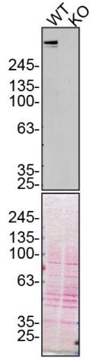
- Western blot analysis of Plectin was performed by loading 10 µg of WT (lane 1) and PLEC CRISPR KO (lane 2) U2OS cell lysates in RIPA buffer onto a 4-15% gradient polyacrylamide gel. Proteins were transferred to nitrocellulose membrane and blocked in 5% milk. Ponceau stained transfer of blot is shown. PLEC was detected above 245 kDa using a Plectin polyclonal antibody (Product # PA5-56292) at a dilution of 1:10,000 in 5% BSA in TBST overnight at 4 deg, followed by secondary antibody diluted to 0.2 µg/mL using Goat anti-Rabbit IgG (H+L) HRP antibody (Product # 65-6120). Chemiluminescent detection was performed using Pierce ECL Western Blotting Substrate (Product # 32106). Data courtesy of YCharOS Inc., an open science company with the mission of characterizing commercially available antibodies using knockout validation.
Supportive validation
- Submitted by
- Invitrogen Antibodies (provider)
- Main image

- Experimental details
- Immunofluorescent staining of Plectin in human cell line U-251 MG shows positivity in cytoplasm, intermediate filaments & focal adhesion sites. Samples were probed using a Plectin Polyclonal Antibody (Product # PA5-56292).

Supportive validation
- Submitted by
- Invitrogen Antibodies (provider)
- Main image

- Experimental details
- Immunohistochemical staining of Plectin in human liver using Plectin Polyclonal Antibody (Product # PA5-56292).

- Submitted by
- Invitrogen Antibodies (provider)
- Main image
- Experimental details
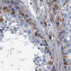
- Immunohistochemical staining of Plectin in human testis using Plectin Polyclonal Antibody (Product # PA5-56292).

- Submitted by
- Invitrogen Antibodies (provider)
- Main image
- Experimental details
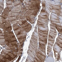
- Immunohistochemical staining of Plectin in human skeletal muscle using Plectin Polyclonal Antibody (Product # PA5-56292) shows high expression.

- Submitted by
- Invitrogen Antibodies (provider)
- Main image
- Experimental details
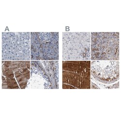
- Immunohistochemical staining of Plectin in human liver, pancreas, skeletal muscle and testis using Plectin Polyclonal Antibody (Product # PA5-56292) (A) shows similar protein distribution across tissues to an independent Plectin Polyclonal Antibody (B).

- Submitted by
- Invitrogen Antibodies (provider)
- Main image
- Experimental details
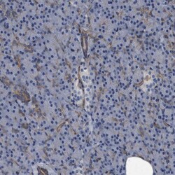
- Immunohistochemical staining of Plectin in human pancreas using Plectin Polyclonal Antibody (Product # PA5-56292) shows low expression as expected.

Supportive validation
- Submitted by
- Invitrogen Antibodies (provider)
- Main image
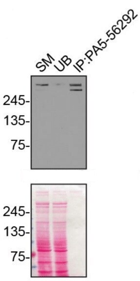
- Experimental details
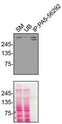
- Immunoprecipitation of Plectin was performed on U2OS cell lysates. Antibody-bead conjugates were prepared by adding 1 µg of PLEC polyclonal antibody (Product # PA5-56292) with 30 µL of protein A -Sepharose beads and rocked overnight at 4°C. 1 µg of PLEC KO lysate was incubated with antibody-bead conjugate for 2 hrs at 4°C. After multiple washes, 10% starting material (SM), 10% unbound fraction (UB) and immunoprecipitated fraction (IP) were processed for immunoblot using a PLEC monoclonal antibody. Ponceau stained transfer of blot is shown. Data courtesy of YCharOS Inc., an open science company with the mission of characterizing commercially available antibodies using knockout validation.