Explore
 Validate
 Learn
Learn Western blot
Antibody data
- Antibody Data
- Antigen structure
- References [0]
- Comments [0]
- Validations
- Western blot [1]
- Immunocytochemistry [1]
- Immunohistochemistry [2]
Submit
Validation data
Reference
Comment
Report error
- Product number
- MA5-32102 - Provider product page

- Provider
- Invitrogen Antibodies
- Product name
- Plectin Recombinant Rabbit Monoclonal Antibody (SY29-04)
- Antibody type
- Monoclonal
- Antigen
- Recombinant full-length protein
- Reactivity
- Human, Mouse, Rat
- Host
- Rabbit
- Isotype
- IgG
- Antibody clone number
- SY29-04
- Vial size
- 100 µL
- Concentration
- 1 mg/mL
- Storage
- Store at 4°C short term. For long term storage, store at -20°C, avoiding freeze/thaw cycles.
No comments: Submit comment
Supportive validation
- Submitted by
- Invitrogen Antibodies (provider)
- Main image
- Experimental details
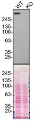
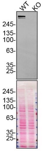
- Western blot analysis of Plectin was performed by loading 10 µg of WT (lane 1) and PLEC CRISPR KO (lane 2) U2OS cell lysates in RIPA buffer onto a 4-15% gradient polyacrylamide gel. Proteins were transferred to nitrocellulose membrane and blocked in 5% milk. Ponceau stained transfer of blot is shown. Plectin was detected above 245 kDa using a Plectin recombinant monoclonal antibody (Product # MA5-32102) at a dilution of 1:5,000 in 5% BSA in TBST overnight at 4 deg, followed by secondary antibody diluted to 0.2 µg/mL using Goat anti-Rabbit IgG (H+L) HRP antibody (Product # 65-6120). Chemiluminescent detection was performed using Pierce ECL Western Blotting Substrate (Product # 32106). Data courtesy of YCharOS Inc., an open science company with the mission of characterizing commercially available antibodies using knockout validation.
Supportive validation
- Submitted by
- Invitrogen Antibodies (provider)
- Main image
- Experimental details
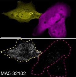
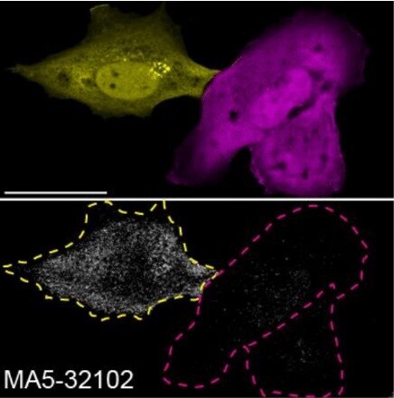
- Immunofluorescence of Plectin was performed using parental and Plectin CRISPR KO cells that were transfected with a GFP or mCherry plasmid, respectively. At 48 hrs post transfection parental and KO cells were mixed and plated to a 1:1 ratio on coverslips as a mosaic and incubated for 24 hrs. Cells were fixed in 4% PFA for 15 min and permeabilized with 0.1% Triton X-100. Cells were stained with Plectin recombinant monoclonal antibody (Product # MA5-32102) at a 1:1,000 dilution overnight at 4 deg. Secondary antibody incubation was performed using 1 µg/mL of Goat anti-Rabbit IgG (H+L) Highly Cross-Adsorbed Alexa Fluor 647 antibody (Product # A-21245) for 1 hr at RT. Imaging was performed with a 40X oil objective and analysis was performed using Image J. Cell image represents a single focal plane. Data courtesy of YCharOS Inc., an open science company with the mission of characterizing commercially available antibodies using knockout validation.
Supportive validation
- Submitted by
- Invitrogen Antibodies (provider)
- Main image
- Experimental details
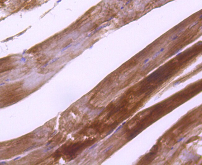
- Immunohistochemical analysis of Plectin of paraffin-embedded rat skeletal muscle tissue using a Plectin Monoclonal antibody (Product #MA5-32102). Counter stained with hematoxylin.

- Submitted by
- Invitrogen Antibodies (provider)
- Main image

- Experimental details
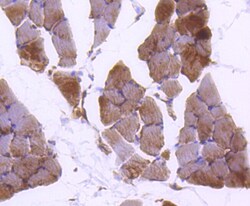
- Immunohistochemical analysis of Plectin of paraffin-embedded Mouse skeletal muscle tissue using a Plectin Monoclonal antibody (Product #MA5-32102). Counter stained with hematoxylin.