Antibody data
- Antibody Data
- Antigen structure
- References [21]
- Comments [0]
- Validations
- Western blot [2]
- Immunocytochemistry [1]
Submit
Validation data
Reference
Comment
Report error
- Product number
- AF1458 - Provider product page

- Provider
- R&D Systems
- Product name
- Human/Mouse/Rat HTRA2/Omi Antibody
- Antibody type
- Polyclonal
- Description
- Immunogen affinity purified. Detects human, mouse, and rat full length and mitochondria-processed HTRA2/Omi.
- Reactivity
- Human, Mouse, Rat
- Host
- Rabbit
- Conjugate
- Unconjugated
- Antigen sequence
O43464- Isotype
- IgG
- Vial size
- 100 ug
- Concentration
- LYOPH
- Storage
- Use a manual defrost freezer and avoid repeated freeze-thaw cycles. 12 months from date of receipt, -20 to -70 °C as supplied. 1 month, 2 to 8 °C under sterile conditions after reconstitution. 6 months, -20 to -70 °C under sterile conditions after reconstitution.
Submitted references PARL mediates Smac proteolytic maturation in mitochondria to promote apoptosis.
Protease Omi cleaving Hax-1 protein contributes to OGD/R-induced mitochondrial damage in neuroblastoma N2a cells and cerebral injury in MCAO mice.
Heat shock inhibition of CDK5 increases NOXA levels through miR-23a repression.
PINK1 kinase catalytic activity is regulated by phosphorylation on serines 228 and 402.
Protease Omi facilitates neurite outgrowth in mouse neuroblastoma N2a cells by cleaving transcription factor E2F1.
Neural-specific deletion of Htra2 causes cerebellar neurodegeneration and defective processing of mitochondrial OPA1.
p53-mediated activation of the mitochondrial protease HtrA2/Omi prevents cell invasion.
Influenza A virus protein PB1-F2 translocates into mitochondria via Tom40 channels and impairs innate immunity.
The ubiquitin-conjugating enzymes UBE2N, UBE2L3 and UBE2D2/3 are essential for Parkin-dependent mitophagy.
PINK1 is degraded through the N-end rule pathway.
The accumulation of misfolded proteins in the mitochondrial matrix is sensed by PINK1 to induce PARK2/Parkin-mediated mitophagy of polarized mitochondria.
PINK1/Parkin-mediated mitophagy is dependent on VDAC1 and p62/SQSTM1.
Novel mitochondrial substrates of omi indicate a new regulatory role in neurodegenerative disorders.
Enhanced HtrA2/Omi expression in oxidative injury to retinal pigment epithelial cells and murine models of neurodegeneration.
Identification of a novel protein MICS1 that is involved in maintenance of mitochondrial morphology and apoptotic release of cytochrome c.
HtrA2 regulates beta-amyloid precursor protein (APP) metabolism through endoplasmic reticulum-associated degradation.
Role and regulation of nodal/activin receptor-like kinase 7 signaling pathway in the control of ovarian follicular atresia.
Yersinia YopP-induced apoptotic cell death in murine dendritic cells is partially independent from action of caspases and exhibits necrosis-like features.
Induction of BIM(EL) following growth factor withdrawal is a key event in caspase-dependent apoptosis of 661W photoreceptor cells.
Roscovitine-induced up-regulation of p53AIP1 protein precedes the onset of apoptosis in human MCF-7 breast cancer cells.
Motoneuron resistance to apoptotic cell death in vivo correlates with the ratio between X-linked inhibitor of apoptosis proteins (XIAPs) and its inhibitor, XIAP-associated factor 1.
Saita S, Nolte H, Fiedler KU, Kashkar H, Venne AS, Zahedi RP, Krüger M, Langer T
Nature cell biology 2017 Apr;19(4):318-328
Nature cell biology 2017 Apr;19(4):318-328
Protease Omi cleaving Hax-1 protein contributes to OGD/R-induced mitochondrial damage in neuroblastoma N2a cells and cerebral injury in MCAO mice.
Wu JY, Li M, Cao LJ, Sun ML, Chen D, Ren HG, Xia Q, Tao ZT, Qin ZH, Hu QS, Wang GH
Acta pharmacologica Sinica 2015 Sep;36(9):1043-52
Acta pharmacologica Sinica 2015 Sep;36(9):1043-52
Heat shock inhibition of CDK5 increases NOXA levels through miR-23a repression.
Morey TM, Roufayel R, Johnston DS, Fletcher AS, Mosser DD
The Journal of biological chemistry 2015 May 1;290(18):11443-54
The Journal of biological chemistry 2015 May 1;290(18):11443-54
PINK1 kinase catalytic activity is regulated by phosphorylation on serines 228 and 402.
Aerts L, Craessaerts K, De Strooper B, Morais VA
The Journal of biological chemistry 2015 Jan 30;290(5):2798-811
The Journal of biological chemistry 2015 Jan 30;290(5):2798-811
Protease Omi facilitates neurite outgrowth in mouse neuroblastoma N2a cells by cleaving transcription factor E2F1.
Ma Q, Hu QS, Xu RJ, Zhen XC, Wang GH
Acta pharmacologica Sinica 2015 Aug;36(8):966-75
Acta pharmacologica Sinica 2015 Aug;36(8):966-75
Neural-specific deletion of Htra2 causes cerebellar neurodegeneration and defective processing of mitochondrial OPA1.
Patterson VL, Zullo AJ, Koenig C, Stoessel S, Jo H, Liu X, Han J, Choi M, DeWan AT, Thomas JL, Kuan CY, Hoh J
PloS one 2014;9(12):e115789
PloS one 2014;9(12):e115789
p53-mediated activation of the mitochondrial protease HtrA2/Omi prevents cell invasion.
Yamauchi S, Hou YY, Guo AK, Hirata H, Nakajima W, Yip AK, Yu CH, Harada I, Chiam KH, Sawada Y, Tanaka N, Kawauchi K
The Journal of cell biology 2014 Mar 31;204(7):1191-207
The Journal of cell biology 2014 Mar 31;204(7):1191-207
Influenza A virus protein PB1-F2 translocates into mitochondria via Tom40 channels and impairs innate immunity.
Yoshizumi T, Ichinohe T, Sasaki O, Otera H, Kawabata S, Mihara K, Koshiba T
Nature communications 2014 Aug 20;5:4713
Nature communications 2014 Aug 20;5:4713
The ubiquitin-conjugating enzymes UBE2N, UBE2L3 and UBE2D2/3 are essential for Parkin-dependent mitophagy.
Geisler S, Vollmer S, Golombek S, Kahle PJ
Journal of cell science 2014 Aug 1;127(Pt 15):3280-93
Journal of cell science 2014 Aug 1;127(Pt 15):3280-93
PINK1 is degraded through the N-end rule pathway.
Yamano K, Youle RJ
Autophagy 2013 Nov 1;9(11):1758-69
Autophagy 2013 Nov 1;9(11):1758-69
The accumulation of misfolded proteins in the mitochondrial matrix is sensed by PINK1 to induce PARK2/Parkin-mediated mitophagy of polarized mitochondria.
Jin SM, Youle RJ
Autophagy 2013 Nov 1;9(11):1750-7
Autophagy 2013 Nov 1;9(11):1750-7
PINK1/Parkin-mediated mitophagy is dependent on VDAC1 and p62/SQSTM1.
Geisler S, Holmström KM, Skujat D, Fiesel FC, Rothfuss OC, Kahle PJ, Springer W
Nature cell biology 2010 Feb;12(2):119-31
Nature cell biology 2010 Feb;12(2):119-31
Novel mitochondrial substrates of omi indicate a new regulatory role in neurodegenerative disorders.
Johnson F, Kaplitt MG
PloS one 2009 Sep 18;4(9):e7100
PloS one 2009 Sep 18;4(9):e7100
Enhanced HtrA2/Omi expression in oxidative injury to retinal pigment epithelial cells and murine models of neurodegeneration.
Ding X, Patel M, Shen D, Herzlich AA, Cao X, Villasmil R, Klupsch K, Tuo J, Downward J, Chan CC
Investigative ophthalmology & visual science 2009 Oct;50(10):4957-66
Investigative ophthalmology & visual science 2009 Oct;50(10):4957-66
Identification of a novel protein MICS1 that is involved in maintenance of mitochondrial morphology and apoptotic release of cytochrome c.
Oka T, Sayano T, Tamai S, Yokota S, Kato H, Fujii G, Mihara K
Molecular biology of the cell 2008 Jun;19(6):2597-608
Molecular biology of the cell 2008 Jun;19(6):2597-608
HtrA2 regulates beta-amyloid precursor protein (APP) metabolism through endoplasmic reticulum-associated degradation.
Huttunen HJ, Guénette SY, Peach C, Greco C, Xia W, Kim DY, Barren C, Tanzi RE, Kovacs DM
The Journal of biological chemistry 2007 Sep 21;282(38):28285-95
The Journal of biological chemistry 2007 Sep 21;282(38):28285-95
Role and regulation of nodal/activin receptor-like kinase 7 signaling pathway in the control of ovarian follicular atresia.
Wang H, Jiang JY, Zhu C, Peng C, Tsang BK
Molecular endocrinology (Baltimore, Md.) 2006 Oct;20(10):2469-82
Molecular endocrinology (Baltimore, Md.) 2006 Oct;20(10):2469-82
Yersinia YopP-induced apoptotic cell death in murine dendritic cells is partially independent from action of caspases and exhibits necrosis-like features.
Gröbner S, Autenrieth SE, Soldanova I, Gunst DS, Schaller M, Bohn E, Müller S, Leverkus M, Wesselborg S, Autenrieth IB, Borgmann S
Apoptosis : an international journal on programmed cell death 2006 Nov;11(11):1959-68
Apoptosis : an international journal on programmed cell death 2006 Nov;11(11):1959-68
Induction of BIM(EL) following growth factor withdrawal is a key event in caspase-dependent apoptosis of 661W photoreceptor cells.
Gómez-Vicente V, Doonan F, Donovan M, Cotter TG
The European journal of neuroscience 2006 Aug;24(4):981-90
The European journal of neuroscience 2006 Aug;24(4):981-90
Roscovitine-induced up-regulation of p53AIP1 protein precedes the onset of apoptosis in human MCF-7 breast cancer cells.
Wesierska-Gadek J, Gueorguieva M, Horky M
Molecular cancer therapeutics 2005 Jan;4(1):113-24
Molecular cancer therapeutics 2005 Jan;4(1):113-24
Motoneuron resistance to apoptotic cell death in vivo correlates with the ratio between X-linked inhibitor of apoptosis proteins (XIAPs) and its inhibitor, XIAP-associated factor 1.
Perrelet D, Perrin FE, Liston P, Korneluk RG, MacKenzie A, Ferrer-Alcon M, Kato AC
The Journal of neuroscience : the official journal of the Society for Neuroscience 2004 Apr 14;24(15):3777-85
The Journal of neuroscience : the official journal of the Society for Neuroscience 2004 Apr 14;24(15):3777-85
No comments: Submit comment
Supportive validation
- Submitted by
- R&D Systems (provider)
- Main image

- Experimental details
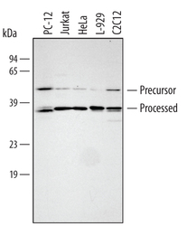
- Detection of Human/Mouse/Rat HTRA2/Omi by Western Blot. Western blot shows lysates of PC-12 rat adrenal pheochromocytoma cell line, Jurkat human acute T cell leukemia cell line, HeLa human cervical epithelial carcinoma cell line, L-929 mouse fibroblast cell line, and C2C12 mouse myoblast cell line. PVDF membrane was probed with 0.25 µg/mL of Rabbit Anti-Human/Mouse/Rat HTRA2/Omi Antigen Affinity-purified Polyclonal Antibody (Catalog # AF1458) followed by HRP-conjugated Anti-Rabbit IgG Secondary Antibody (Catalog # HAF008). Specific bands were detected for HTRA2/Omi at approximately 36 and 49 kDa (as indicated). This experiment was conducted under reducing conditions and using Immunoblot Buffer Group 2.
- Submitted by
- R&D Systems (provider)
- Main image

- Experimental details
- Detection of Human and Mouse HTRA2/Omi by Simple WesternTM. Simple Western lane view shows lysates of C2C12 mouse myoblast cell line and HeLa human cervical epithelial carcinoma cell line, loaded at 0.2 mg/mL. Specific bands were detected for HTRA2/Omi at approximately 51 kDa (precursor) and 41 kDa (processed) (as indicated) using 2.5 µg/mL of Rabbit Anti-Human/Mouse/Rat HTRA2/Omi Antigen Affinity-purified Polyclonal Antibody (Catalog # AF1458). This experiment was conducted under reducing conditions and using the 12-230 kDa separation system.
Supportive validation
- Submitted by
- R&D Systems (provider)
- Main image

- Experimental details
- HTRA2/Omi in Jurkat Human Cell Line. HTRA2/Omi was detected in immersion fixed Jurkat human acute T cell leukemia cell line stimulated with staurosporin using Rabbit Anti-Human/Mouse/Rat HTRA2/Omi Antigen Affinity-purified Polyclonal Antibody (Catalog # AF1458) at 10 µg/mL for 3 hours at room temperature. Cells were stained using the NorthernLights™ 557-conjugated Anti-Rabbit IgG Secondary Antibody (yellow; Catalog # NL004) and counterstained with DAPI (blue). View our protocol for Fluorescent ICC Staining of Cells on Coverslips.
 Explore
 Validate
 Learn
Learn Western blot