Explore
 Validate
 Learn
Learn Western blot
Antibody data
- Antibody Data
- Antigen structure
- References [0]
- Comments [0]
- Validations
- Western blot [2]
- Immunocytochemistry [1]
- Immunohistochemistry [6]
- Flow cytometry [1]
Submit
Validation data
Reference
Comment
Report error
- Product number
- R32246 - Provider product page

- Provider
- NSJ Bioreagents
- Product name
- BAK Antibody
- Antibody type
- Polyclonal
- Antigen
- Amino acids 22-211 of human BAK were used as the immunogen for the BAK antibody.
- Description
- Antigen affinity
- Reactivity
- Human, Mouse, Rat
- Host
- Rabbit
- Conjugate
- Unconjugated
- Vial size
- 100 µg
- Concentration
- Lyophilized; resuspend with 200 ul for 0.5 mg/ml
- Storage
- After reconstitution, the BAK antibody can be stored for up to one month at 4oC. For long-term, aliquot and store at -20oC. Avoid repeated freezing and thawing.
No comments: Submit comment
Supportive validation
- Submitted by
- NSJ Bioreagents (provider)
- Main image

- Experimental details
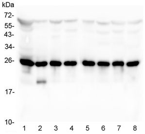
- Western blot testing of human 1) PC-3, 2) HEK293, 3) Jurkat, 4) Caco-2, 5) U-2 OS, 6) HepG2, 7) HeLa and 8) A549 lysate with BAK antibody. Expected molecular weight ~23 kDa.
- Submitted by
- NSJ Bioreagents (provider)
- Main image

- Experimental details
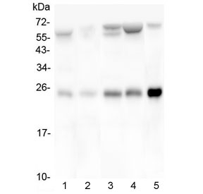
- Western blot testing of 1) rat skeletal muscle, 2) rat lung, 3) mouse heart, 4) mouse skeletal muscle and 5) mouse lung lysate with BAK antibody. Expected molecular weight ~23 kDa.
Supportive validation
- Submitted by
- NSJ Bioreagents (provider)
- Main image

- Experimental details
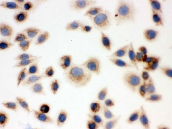
- ICC testing of FFPE human SMMC-7721 cells with BAK antibody. HIER: Boil the paraffin sections in pH 6, 10mM citrate buffer for 20 minutes and allow to cool prior to staining.
Supportive validation
- Submitted by
- NSJ Bioreagents (provider)
- Main image

- Experimental details
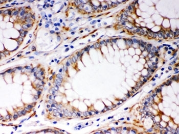
- IHC testing of FFPE human intestinal cancer tissue with BAK antibody. HIER: Boil the paraffin sections in pH 6, 10mM citrate buffer for 20 minutes and allow to cool prior to staining.
- Submitted by
- NSJ Bioreagents (provider)
- Main image

- Experimental details
- IHC testing of FFPE mouse skeletal muscle with BAK antibody. HIER: Boil the paraffin sections in pH 6, 10mM citrate buffer for 20 minutes and allow to cool prior to staining.

- Submitted by
- NSJ Bioreagents (provider)
- Main image

- Experimental details
- IHC testing of FFPE rat skeletal muscle with BAK antibody. HIER: Boil the paraffin sections in pH 6, 10mM citrate buffer for 20 minutes and allow to cool prior to staining.
- Submitted by
- NSJ Bioreagents (provider)
- Main image

- Experimental details
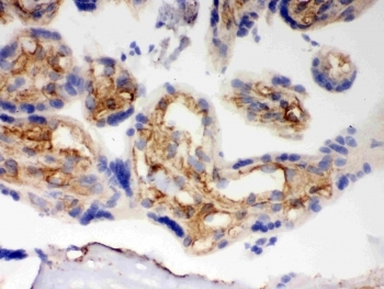
- IHC testing of frozen human placental tissue with BAK antibody.
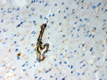
- Submitted by
- NSJ Bioreagents (provider)
- Main image
- Experimental details
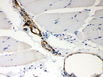
- IHC testing of frozen mouse heart tissue with BAK antibody.
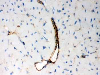
- Submitted by
- NSJ Bioreagents (provider)
- Main image

- Experimental details
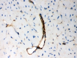
- IHC testing of frozen rat heart tissue with BAK antibody.
Supportive validation
- Submitted by
- NSJ Bioreagents (provider)
- Main image

- Experimental details
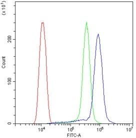
- Flow cytometry testing of human ThP1 cells with BAK antibody at 1ug/10^6 cells (blocked with goat sera); Red=cells alone, Green=isotype control, Blue= BAK antibody.