Explore
 Validate
 Learn
Learn Western blot
 Immunocytochemistry
Antibody data
- Antibody Data
- Antigen structure
- References [1]
- Comments [0]
- Validations
- Western blot [1]
- Immunohistochemistry [2]
- Flow cytometry [1]
Submit
Validation data
Reference
Comment
Report error
- Product number
- NBP2-67460 - Provider product page

- Provider
- Novus Biologicals
- Product name
- Rabbit Monoclonal BAK Antibody
- Antibody type
- Monoclonal
- Description
- Protein A purified.
- Reactivity
- Human, Mouse
- Host
- Rabbit
- Isotype
- IgG
- Vial size
- 100 ul
- Storage
- Store at 4C short term. Aliquot and store at -20C long term. Avoid freeze-thaw cycles.
Submitted references YWHAZ amplification/overexpression defines aggressive bladder cancer and contributes to chemo-/radio-resistance by suppressing caspase-mediated apoptosis.
Yu CC, Li CF, Chen IH, Lai MT, Lin ZJ, Korla PK, Chai CY, Ko G, Chen CM, Hwang T, Lee SC, Sheu JJ
The Journal of pathology 2019 Aug;248(4):476-487
The Journal of pathology 2019 Aug;248(4):476-487
No comments: Submit comment
Supportive validation
- Submitted by
- Novus Biologicals (provider)
- Main image

- Experimental details
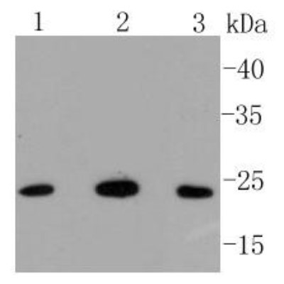
- Western Blot: BAK Antibody (SU32-07) [NBP2-67460] - Analysis of Bak on different lysates using anti-Bak antibody at 1/1,000 dilution. Positive control: Lane 1: Hela Lane 2: Human skeletal muscle Lane 3: Ags
Supportive validation
- Submitted by
- Novus Biologicals (provider)
- Main image

- Experimental details
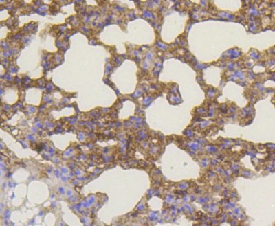
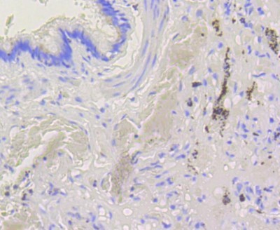
- Immunohistochemistry-Paraffin: BAK Antibody (SU32-07) [NBP2-67460] - Analysis of paraffin-embedded mouse lung tissue using anti-Bak antibody. Counter stained with hematoxylin.
- Submitted by
- Novus Biologicals (provider)
- Main image
- Experimental details
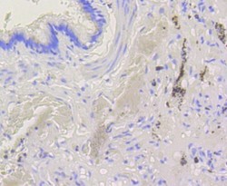
- Immunohistochemistry-Paraffin: BAK Antibody (SU32-07) [NBP2-67460] - Analysis of paraffin-embedded human lung tissue using anti-Bak antibody. Counter stained with hematoxylin.
Supportive validation
- Submitted by
- Novus Biologicals (provider)
- Main image
- Experimental details
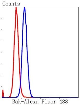
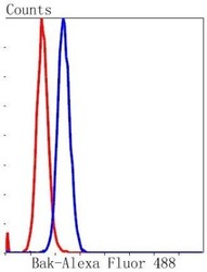
- Flow Cytometry: BAK Antibody (SU32-07) [NBP2-67460] - Analysis of NIH/3T3 cells with Bak antibody at 1/50 dilution (blue) compared with an unlabelled control (cells without incubation with primary antibody; red). Alexa Fluor 488-conjugated goat anti rabbit IgG was used as the secondary antibody.