Explore
 Validate
 Learn
Learn Western blot
 ELISA
ELISAAntibody data
- Antibody Data
- Antigen structure
- References [0]
- Comments [0]
- Validations
- Western blot [1]
- Flow cytometry [3]
Submit
Validation data
Reference
Comment
Report error
- Product number
- NBP1-74026-100ul - Provider product page

- Provider
- Novus Biologicals
- Proper citation
- Novus Cat#NBP1-74026-100ul, RRID:AB_11139509
- Product name
- Mouse Monoclonal BAK Antibody
- Antibody type
- Monoclonal
- Description
- Protein G purified.
- Reactivity
- Human
- Host
- Mouse
- Isotype
- IgG
- Vial size
- 100 ul
- Concentration
- 1 mg/ml
- Storage
- Store at 4C short term. Aliquot and store at -20C long term. Avoid freeze-thaw cycles.
No comments: Submit comment
Supportive validation
- Submitted by
- Novus Biologicals (provider)
- Main image

- Experimental details
- Western Blot: BAK Antibody (AT38E2) [NBP1-74026] - The cell lysate (40ug) were resolved by SDS-PAGE, transferred to PVDF membrane and probed with anti-human BAK1 antibody (1:1000). Proteins were visualized using a goat anti-mouse secondary antibody conjugated to HRP and an ECL detection system. Lane 1 : 293T cell lysate Lane 2 : HeLa cell lysate Lane 3 : A431 cell lysate Lane 4 : A549 cell lysate Lane 5 : Jurkat cell lysate Lane 6 : MCF7 cell lysate Lane 7 : PC3 cell lysate

Supportive validation
- Submitted by
- Novus Biologicals (provider)
- Main image

- Experimental details
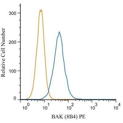
- Flow Cytometry: BAK Antibody (AT38E2) [NBP1-74026] - Flow Cytometry: BAK Antibody (8B4) [PE] [NBP1-74026PE] - An intracellular stain was performed on RAW 246.7 cells with BAK antibody (8B4) NBP1-74026PE (blue) and a matched isotype control NB600-985PE (orange). Cells were fixed with 4% PFA and then permeablized with 0.1% saponin. Cells were incubated in an antibody dilution of 1 ug/mL for 30 minutes at room temperature. Both antibodies were conjugated to phycoerythrin. Image using the PE form of this antibody.
- Submitted by
- Novus Biologicals (provider)
- Main image

- Experimental details
- Flow Cytometry: BAK Antibody (AT38E2) [NBP1-74026] - Flow Cytometry: BAK Antibody (8B4) [PE] [NBP1-74026PE] - An intracellular stain was performed on Jurkat cells with BAK antibody (8B4) NBP1-74026PE (blue) and a matched isotype control NB600-985PE (orange). Cells were fixed with 4% PFA and then permeablized with 0.1% saponin. Cells were incubated in an antibody dilution of 1 ug/mL for 30 minutes at room temperature. Both antibodies were conjugated to phycoerythrin. Image using the PE form of this antibody.

- Submitted by
- Novus Biologicals (provider)
- Main image

- Experimental details
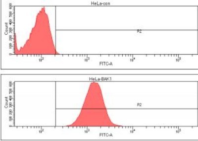
- Flow Cytometry: BAK Antibody (AT38E2) [NBP1-74026] - Analysis of BAK1 in HeLa cell line, staining at 2-5ug for 1x10⁶cells (red line). The secondary antibody used goat anti-mouse IgG Alexa fluor 488 conjugate. Isotype control antibody was mouse IgG (black line).