 Explore
 Validate
 Learn
Learn Western blot
 ELISA
ELISAAntibody data
- Antibody Data
- Antigen structure
- References [0]
- Comments [0]
- Validations
- Western blot [5]
- Immunocytochemistry [1]
- Immunohistochemistry [4]
- Flow cytometry [1]
Submit
Validation data
Reference
Comment
Report error
- Product number
- RQ4439 - Provider product page

- Provider
- NSJ Bioreagents
- Product name
- p65 Antibody / NF-kB
- Antibody type
- Polyclonal
- Description
- Antigen affinity purified
- Reactivity
- Human, Mouse, Rat
- Host
- Rabbit
- Conjugate
- Unconjugated
- Vial size
- 100 µg
- Storage
- After reconstitution, the p65 antibody can be stored for up to one month at 4°C. For long-term, aliquot and store at -20deg;C. Avoid repeated freezing and thawing.
No comments: Submit comment
Supportive validation
- Submitted by
- NSJ Bioreagents (provider)
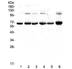
- Main image

- Experimental details
- Western blot testing of human 1) COLO-320, 2) A549, 3) HepG2, 4) MDA-MB-231, 5) PANC-1 and 6) A375 lysate with p65 antibody at 0.5ug/ml. Expected molecular weight ~65 kDa.
- Submitted by
- NSJ Bioreagents (provider)
- Main image

- Experimental details
- Western blot testing of rat 1) spleen, 2) lung, 3) kidney, 4) testis and mouse 5) spleen, 6) lung, 7) testis and 8) NIH3T3 lysate with p65 antibody at 0.5ug/ml. Expected molecular weight ~65 kDa.

- Submitted by
- NSJ Bioreagents (provider)
- Main image

- Experimental details
- Western blot testing of human 1) COLO-320, 2) A549, 3) HepG2, 4) MDA-MB-231, 5) PANC-1 and 6) A375 lysate with p65 antibody at 0.5ug/ml. Expected molecular weight ~65 kDa.

- Submitted by
- NSJ Bioreagents (provider)
- Main image
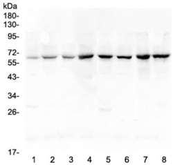
- Experimental details
- Western blot testing of rat 1) spleen, 2) lung, 3) kidney, 4) testis and mouse 5) spleen, 6) lung, 7) testis and 8) NIH3T3 lysate with p65 antibody at 0.5ug/ml. Expected molecular weight ~65 kDa.

- Submitted by
- NSJ Bioreagents (provider)
- Main image

- Experimental details
- Western blot testing of 1) human HeLa, 2) human Raji, 3) human HepG2, 4) human K562, 5) rat PC-12, 6) rat RH35, 7) mouse RAW264.7 and 8) mouse HEPA1-6 cell lysate with p65 antibody at 0.5ug/ml. Expected molecular weight ~65 kDa.

Supportive validation
- Submitted by
- NSJ Bioreagents (provider)
- Main image

- Experimental details
- Immunofluorescent staining of FFPE human U-2 OS cells with p65 antibody (green) and DAPI nuclear stain (blue). HIER: steam section in pH6 citrate buffer for 20 min.

Supportive validation
- Submitted by
- NSJ Bioreagents (provider)
- Main image
- Experimental details
- IHC testing of FFPE human colon cancer with p65 antibody at 2ug/ml. HIER: boil tissue sections in pH6, 10mM citrate buffer, for 10-20 min followed by cooling at RT for 20 min.

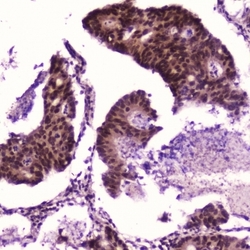
- Submitted by
- NSJ Bioreagents (provider)
- Main image

- Experimental details
- IHC testing of FFPE human breast cancer with p65 antibody at 2ug/ml. HIER: boil tissue sections in pH6, 10mM citrate buffer, for 10-20 min followed by cooling at RT for 20 min.

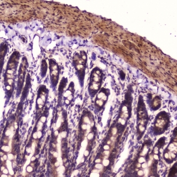
- Submitted by
- NSJ Bioreagents (provider)
- Main image

- Experimental details
- IHC testing of FFPE rat small intestine with p65 antibody at 2ug/ml. HIER: boil tissue sections in pH6, 10mM citrate buffer, for 10-20 min followed by cooling at RT for 20 min.
- Submitted by
- NSJ Bioreagents (provider)
- Main image

- Experimental details
- IHC testing of FFPE rat kidney with p65 antibody at 2ug/ml. HIER: boil tissue sections in pH6, 10mM citrate buffer, for 10-20 min followed by cooling at RT for 20 min.

Supportive validation
- Submitted by
- NSJ Bioreagents (provider)
- Main image
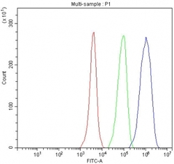
- Experimental details
- Flow cytometry testing of fixed and permeabilized human HeLa cells with p65 antibody at 1ug/million cells (blocked with goat sera); Red=cells alone, Green=isotype control, Blue= p65 antibody.
