Explore
 Validate
 Learn
Learn Western blot
Antibody data
- Antibody Data
- Antigen structure
- References [0]
- Comments [0]
- Validations
- Western blot [2]
- Immunohistochemistry [1]
Submit
Validation data
Reference
Comment
Report error
- Product number
- R1170 - Provider product page

- Provider
- Acris Antibodies GmbH
- Proper citation
- Acris Antibodies GmbH Cat#R1170, RRID:AB_1005683
- Product name
- anti RELA / NF-kB p65 pSer276
- Antibody type
- Polyclonal
- Antigen
- NFkB p65 (Rel A) peptide corresponding to a region near phospho Serine 276 of the Human protein conjugated to Keyhole Limpet Hemocyanin (KLH).
- Reactivity
- Human
- Host
- Rabbit
- Vial size
- 0.1 ml
- Concentration
- 80.0 mg/ml (by refractometry)
No comments: Submit comment
Supportive validation
- Submitted by
- Acris Antibodies GmbH (provider)
- Main image

- Experimental details
- TNF induces phosphorylation of p65 in KBM-5 cells. Cytoplasmic and nuclear protein lysates prepared after 0, 5, 10, 15, 30 and 60 minutes of 0.1 nM TNF treatment of KBM-5 cells shows inducible phosphorylation using phospho specific polyclonal anti-Human pSer276 p65 antibody.Pan reactive anti R1009 p65 antibody was used as Control to show the presence of total p65 in both the cytoplasmic and nuclear extracts. Phosphorylation of p65 occurs after approximately 10 min of TNF exposure. Migration of phosphorylated p65 into the nucleus occurs within a similar time frame. HRP conjugated Goat anti-Rabbit IgG was used to develop the blot using a chemiluminescent detection method. Other detection methods will yield similar results. Personal Communication, Aggarwal BB.

- Submitted by
- Acris Antibodies GmbH (provider)
- Main image

- Experimental details
- pSer276 p65 antibody shows phospho p65 staining in carcinoma cells.Immunoblot of total protein lysates from various human head and neck tumors shows phospho p65 staining in tumor cell lines using phospho specific polyclonal anti-human pS276 p65. Lanes 1-6 contain protein lysates from human squamal carcinoma cell lines.Lane 7 is a protein lysate from a primary culture of human keratinocytes and does not show significant levels of phosphorylated p65.Lane 8 contains protein lysate from ATCC SCC9 cells (also a head and neck squamal carcinoma). Lane 9 contains lysate from EGF-induced human derived A431 cells. Lane 10 (not shown) contains a molecular weight standard. Concurrent staining with anti beta microtubulin (not shown) was used to confirm equal protein loading in all lanes. HRP conjugated Goat anti-Rabbit IgG was used to develop the blot using a chemiluminescent detection method. Other detection methods will yield similar results. Data contributed by Yu, M., NIH, personal communication.

Supportive validation
- Submitted by
- Acris Antibodies GmbH (provider)
- Main image

- Experimental details
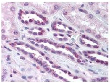
- R1170 pSer276 p65 antibody staining of Formalin-Fixed, Paraffin Embedded Human kidney tissue at 1/500 dilution. No pre-treatment of sample was required. The image shows the localization of antibody as the precipitated red signal, with a hematoxylin purple nuclear counter stain.