Explore
 Validate
 Learn
Learn Western blot
Antibody data
- Antibody Data
- Antigen structure
- References [2]
- Comments [0]
- Validations
- Western blot [5]
- Immunoprecipitation [1]
- Immunohistochemistry [1]
Submit
Validation data
Reference
Comment
Report error
- Product number
- GTX109162 - Provider product page

- Provider
- GeneTex
- Proper citation
- GeneTex Cat#GTX109162, RRID:AB_11163771
- Product name
- 4E-BP1 antibody [N1C3]
- Antibody type
- Polyclonal
- Reactivity
- Human, Rat
- Host
- Rabbit
Submitted references Inflammatory interferon activates HIF-1α-mediated epithelial-to-mesenchymal transition via PI3K/AKT/mTOR pathway.
Aqueous extracts of Paeonia suffruticosa modulates mitochondrial proteostasis by reactive oxygen species-induced endoplasmic reticulum stress in pancreatic cancer cells.
Yeh YH, Hsiao HF, Yeh YC, Chen TW, Li TK
Journal of experimental & clinical cancer research : CR 2018 Mar 27;37(1):70
Journal of experimental & clinical cancer research : CR 2018 Mar 27;37(1):70
Aqueous extracts of Paeonia suffruticosa modulates mitochondrial proteostasis by reactive oxygen species-induced endoplasmic reticulum stress in pancreatic cancer cells.
Liu YH, Weng YP, Tsai HY, Chen CJ, Lee DY, Hsieh CL, Wu YC, Lin JY
Phytomedicine : international journal of phytotherapy and phytopharmacology 2018 Jul 15;46:184-192
Phytomedicine : international journal of phytotherapy and phytopharmacology 2018 Jul 15;46:184-192
No comments: Submit comment
Supportive validation
- Submitted by
- GeneTex (provider)
- Main image
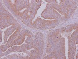
- Experimental details
- 4E-BP1 antibody [N1C3] detects 4E-BP1 protein at cytoplasm on colon carcinoma by immunohistochemical analysis. Sample: Paraffin-embedded colon carcinoma. 4E-BP1 antibody [N1C3] (GTX109162) dilution: 1:500.
- Validation comment
- WB

- Submitted by
- GeneTex (provider)
- Main image

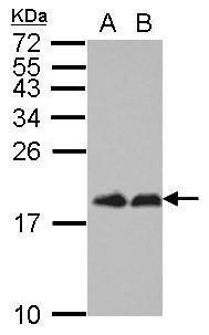
- Experimental details
- Sample (30 ug of whole cell lysate) A: H1299 B: HCT116 15% SDS PAGE GTX109162 diluted at 1:1000
- Validation comment
- WB
- Submitted by
- GeneTex (provider)
- Main image
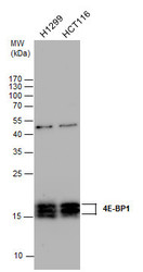
- Experimental details
- 4E-BP1 antibody detects 4E-BP1 protein by western blot analysis. Various whole cell extracts (30 £gg) were separated by 15% SDS-PAGE, and the membrane was blotted with 4E-BP1 antibody (GTX109162) diluted at a dilution of 1:2000.

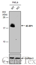
- Submitted by
- GeneTex (provider)
- Main image
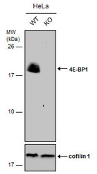
- Experimental details
- Wild-type (WT) and 4E-BP1 knockout (KO) HeLa cell extracts (30 ?g) were separated by 15% SDS-PAGE, and the membrane was blotted with 4E-BP1 antibody [N1C3] (GTX109162) diluted at 1:500. The HRP-conjugated anti-rabbit IgG antibody (GTX213110-01) was used to detect the primary antibody.
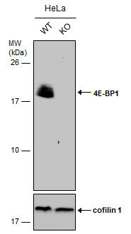
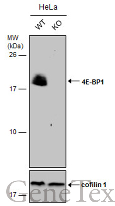
- Submitted by
- GeneTex (provider)
- Main image
- Experimental details
- Wild-type (WT) and 4E-BP1 knockout (KO) HeLa cell extracts (30 ?g) were separated by 15% SDS-PAGE, and the membrane was blotted with 4E-BP1 antibody [N1C3] (GTX109162) diluted at 1:500. The HRP-conjugated anti-rabbit IgG antibody (GTX213110-01) was used to detect the primary antibody.
Supportive validation
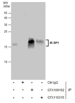
- Submitted by
- GeneTex (provider)
- Main image
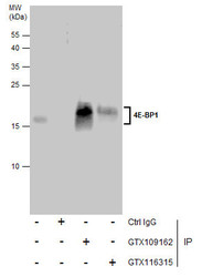
- Experimental details
- Immunoprecipitation of 4E-BP1 protein from HCT116 whole cell extracts using 5 £gg of 4E-BP1 antibody [N1C3] (GTX109162) or 4E-BP1 antibody [N1C3-2] (GTX116315).Western blot analysis was performed using 4E-BP1 antibody [N1C3] (GTX109162).EasyBlot anti-Rabbit IgG (GTX221666-01) was used as a secondary reagent.
Supportive validation
- Submitted by
- GeneTex (provider)
- Main image
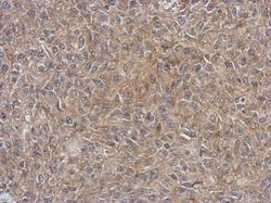
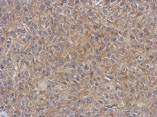
- Experimental details
- Immunohistochemical analysis of paraffin-embedded RT2 xenograft, using EIF4EBP1(GTX109162) antibody at 1:500 dilution.